All SubjectsAnatomy (35)Anesthesiology (9)Biochemistry (6)Community Medicine (14)Dermatology (24)ENT (15)Forensic Medicine (18)General Medicine (22)Internal Medicine (13)Internal Medicine (5)Microbiology (25)Obstetrics and Gynecology (20)Ophthalmology (16)Orthopaedics (11)Pathology (14)Pathology (9)Pediatrics (28)Pharmacology (8)Physiology (14)Radiology (28)Surgery (4)Surgery (15)
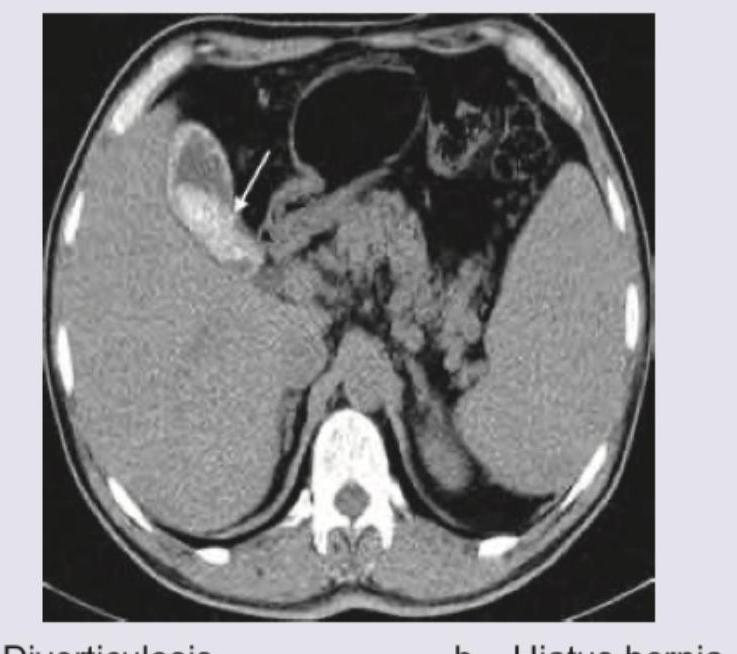
Q21
CT abdomen shows: (Recent NEET Pattern 2016-17)
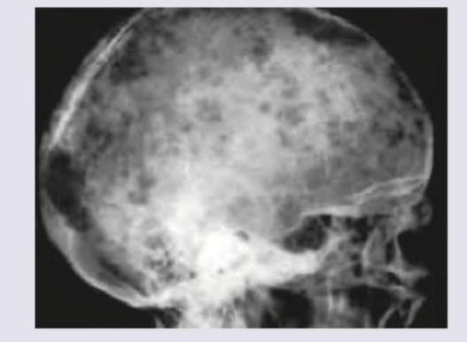
Q22
X-ray skull shows: (Recent NEET Pattern 2016-17)
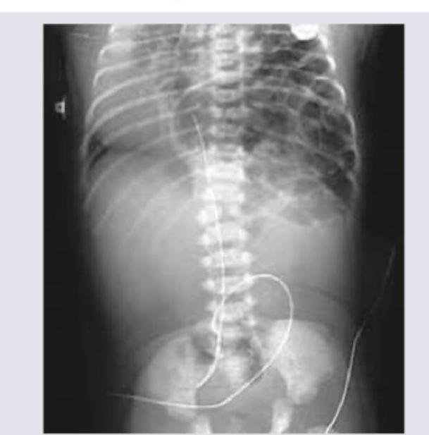
Q23
Identify the condition on the basis of infantogram shown in the image:
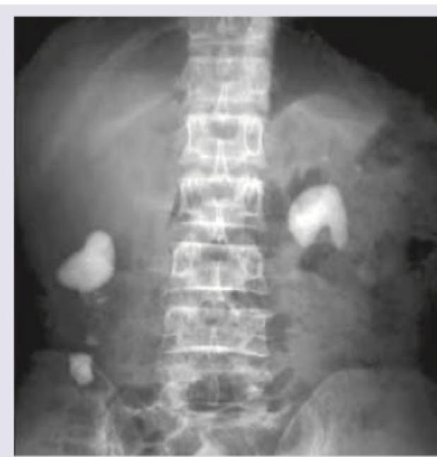
Q24
The given IVU shows:
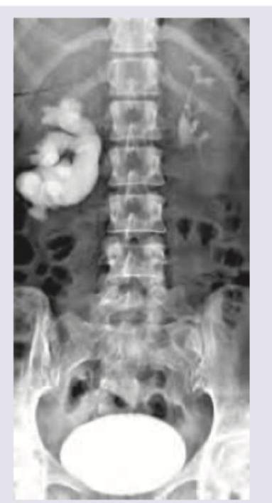
Q25
Which of the following kidney stones best explains the findings in this X-ray KUB?
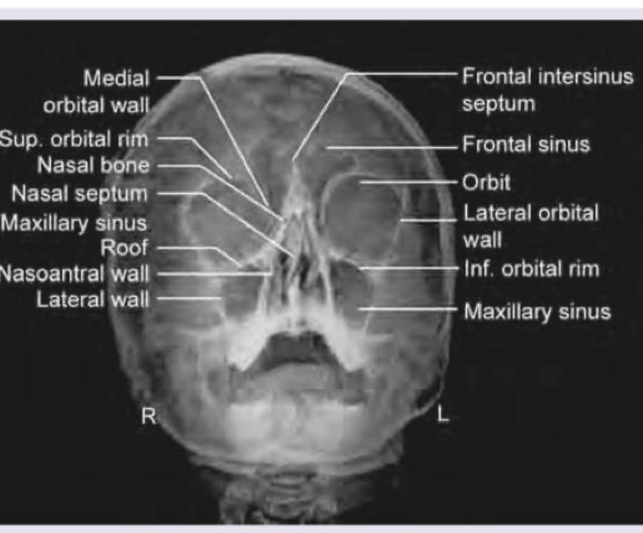
Q26
The given X-ray of paranasal sinuses shows which view?
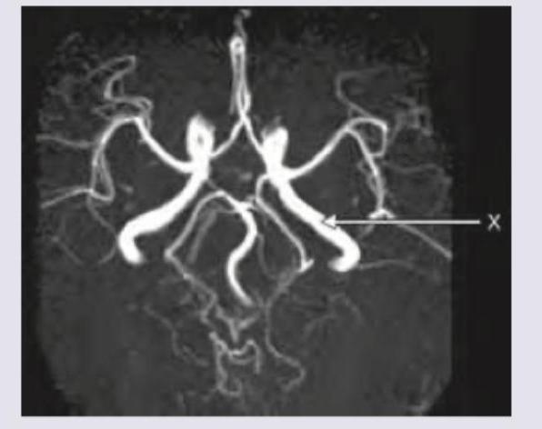
Q27
The blood vessel marked as $X$ in the CT angiography image is:
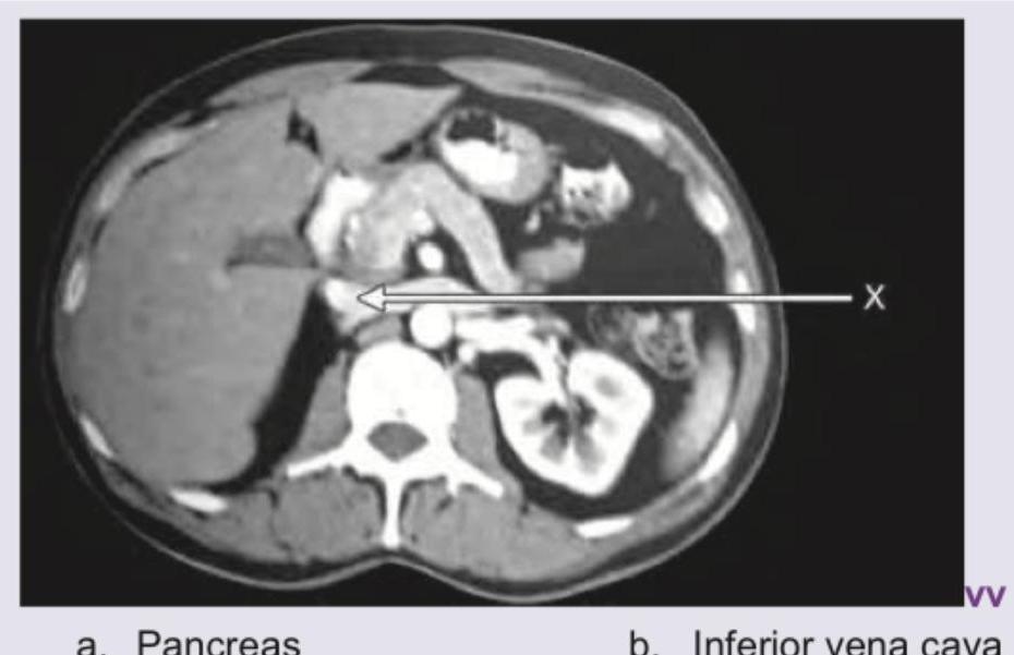
Q28
Name the structure marked as $X$ in the CT abdomen shown below: (Recent NEET Pattern 2016-17)