All SubjectsAnatomy (30)Anesthesiology (8)Biochemistry (8)Community Medicine (17)Dermatology (24)ENT (18)Forensic Medicine (18)General Medicine (2)Internal Medicine (23)Internal Medicine (8)Microbiology (39)Obstetrics and Gynecology (15)Ophthalmology (16)Orthopaedics (11)Pathology (10)Pathology (17)Pediatrics (26)Pharmacology (6)Physiology (15)Radiology (30)Surgery (5)Surgery (22)
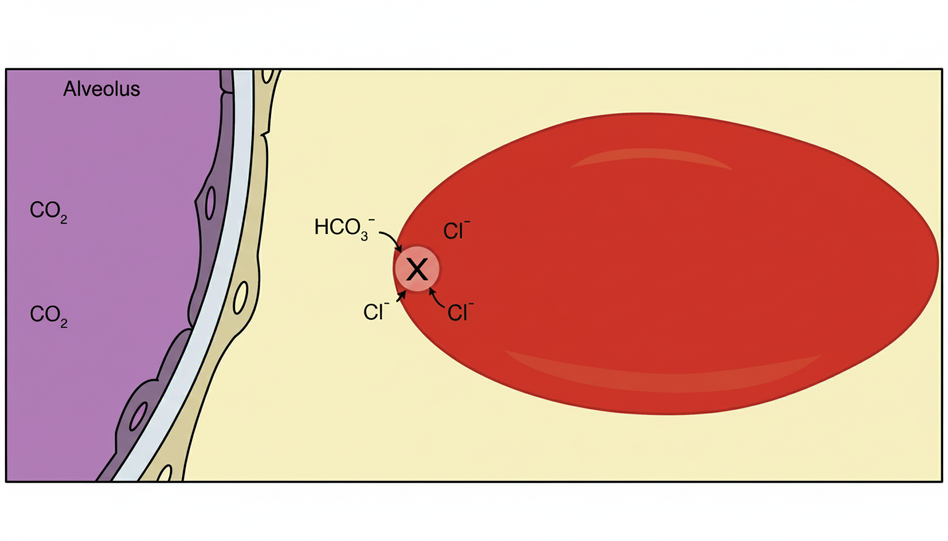
Q11
Which protein is responsible for the effect shown in RBC marked as $X$ ?
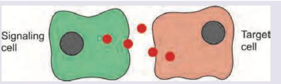
Q12
Identify the modality of intercellular communication shown below.
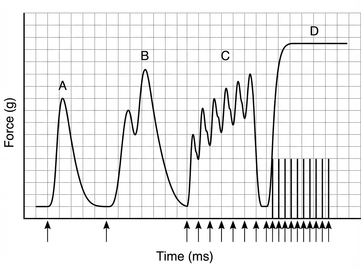
Q13
The following skeletal muscle recording shows presence of: (Recent NEET Pattern 2016-17)
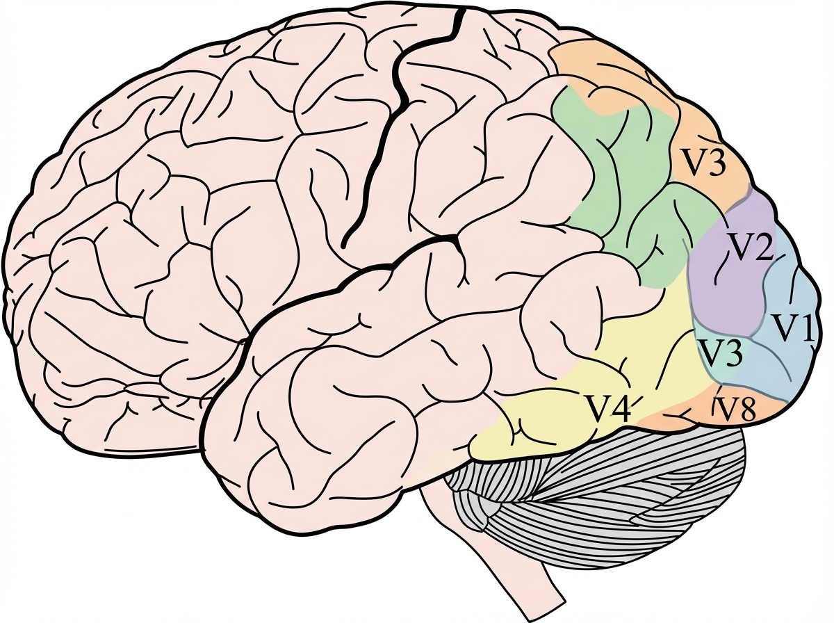
Q14
Which of the following area of visual cortex is related to color vision?
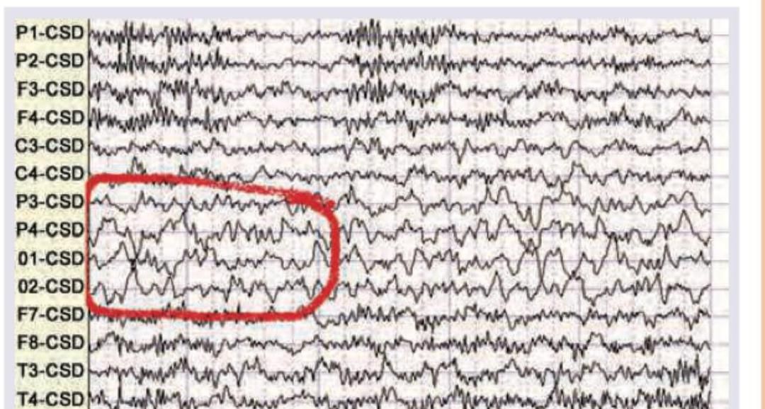
Q15
Which wave is seen in the given EEG recording?