All SubjectsAnatomy (35)Anesthesiology (9)Biochemistry (6)Community Medicine (14)Dermatology (24)ENT (15)Forensic Medicine (18)General Medicine (22)Internal Medicine (13)Internal Medicine (5)Microbiology (25)Obstetrics and Gynecology (20)Ophthalmology (16)Orthopaedics (11)Pathology (14)Pathology (9)Pediatrics (28)Pharmacology (8)Physiology (14)Radiology (28)Surgery (4)Surgery (15)
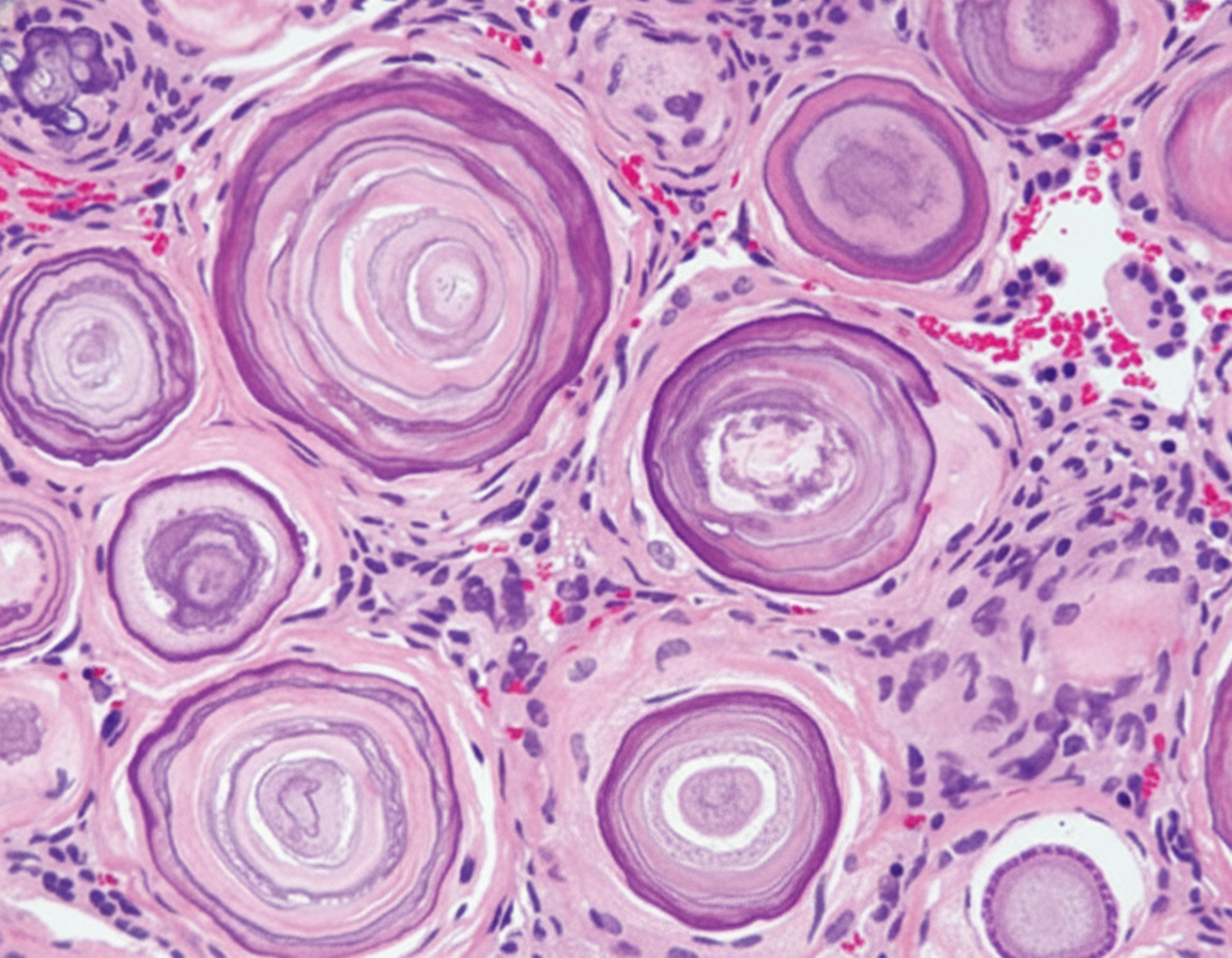
Q11
All are true about the brain tumour associated with the below mentioned histopathological findings except:
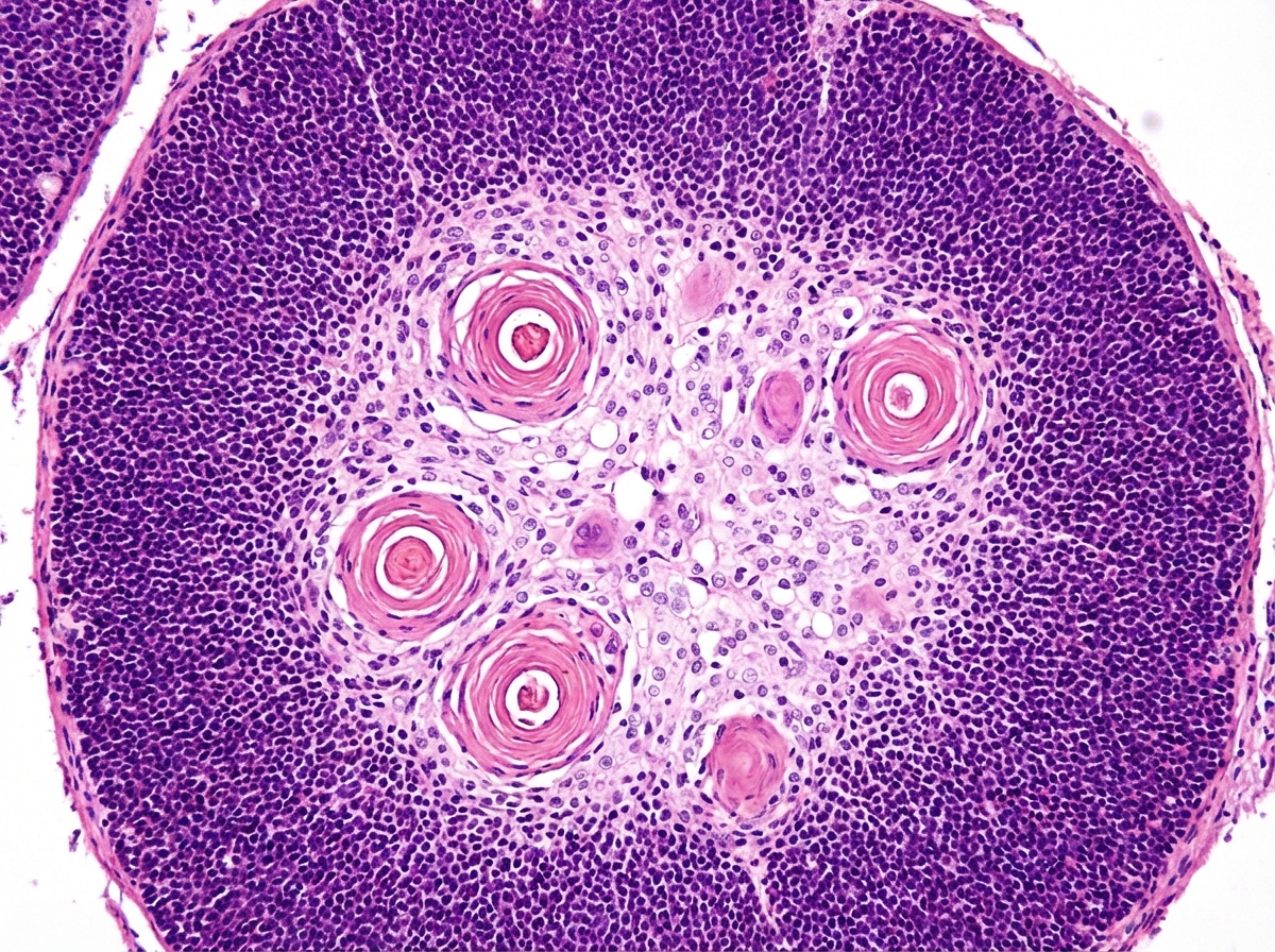
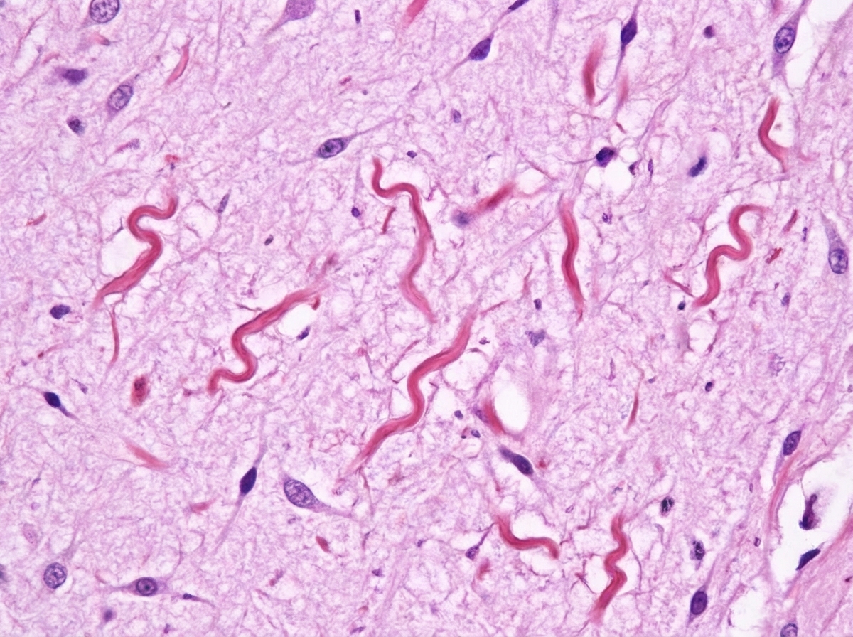
Q12
Cork screw inclusion bodies in brain biopsy specimen are seen in:
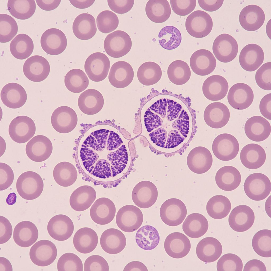
Q13
The image shows: (Recent NEET Pattern 2016-17)
Q14
Identify the tissue: