All (368)Anatomy (30)Anesthesiology (8)Biochemistry (8)Community Medicine (17)Dermatology (24)ENT (18)Forensic Medicine (18)General Medicine (2)Internal Medicine (23)Internal Medicine (8)Microbiology (39)Obstetrics and Gynecology (15)Ophthalmology (16)Orthopaedics (11)Pathology (10)Pathology (17)Pediatrics (26)Pharmacology (6)Physiology (15)Radiology (30)Surgery (5)Surgery (22)
Q41
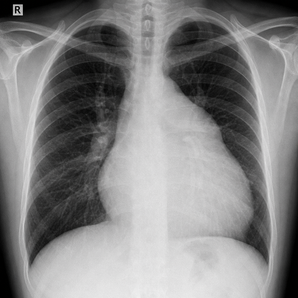
A chest X-ray shows the following appearance. Identify the pathology:
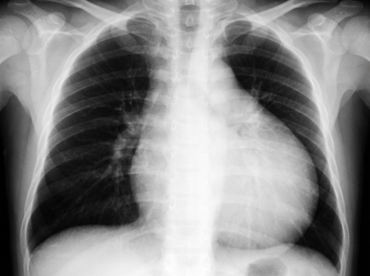
Q42
A 30-year-old hypertension patient presents with daily headaches. The CXR given below shows which of the following? (Recent NEET Pattern 2016-17)
Q43
An adult undergoes multiple FFP transfusions for excessive bleeding after cardiac surgery and develops respiratory distress. CXR done is shown below. What does it indicate?
Q44
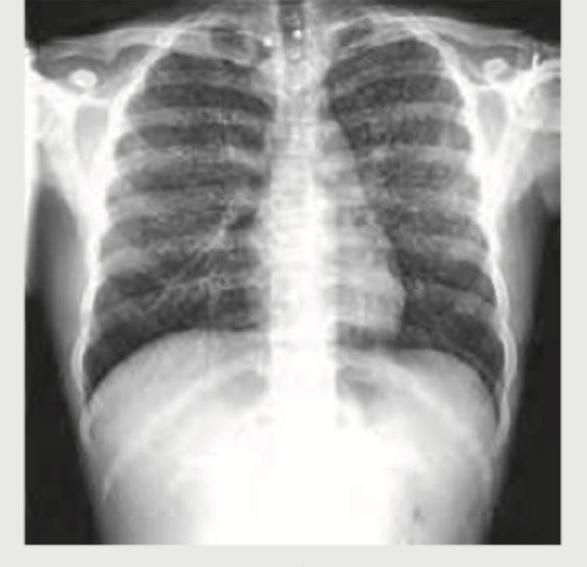
Identify the congenital heart disease presenting with cyanosis in CXR: (Recent NEET Pattern 2016-17)
Q45
A breast cancer patient presents with difficulty in breathing. CXR shows:
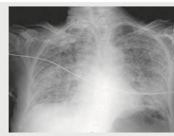
Q46
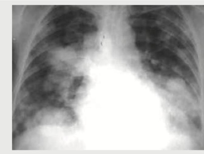
An AIDS patient presents with respiratory distress. CXR shows:
Q47
An 18-year-old boy is brought to the hospital with difficulty in breathing after a bar fight. What does the given CT chest show?
Q48
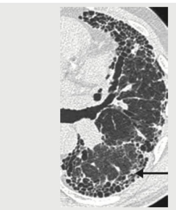
The lung parenchyma on CT chest shown below is best described as:
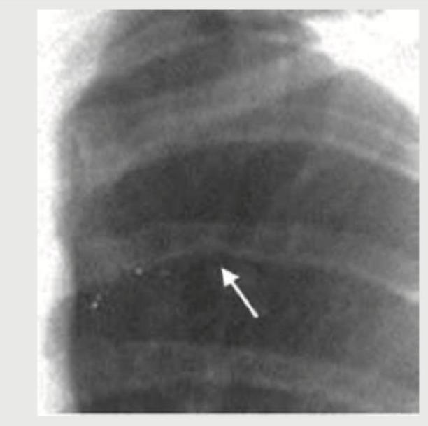
Q49
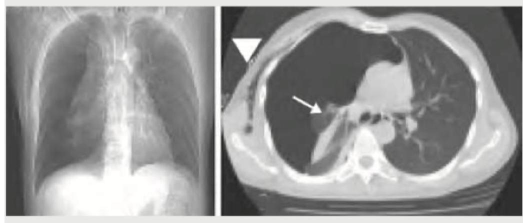
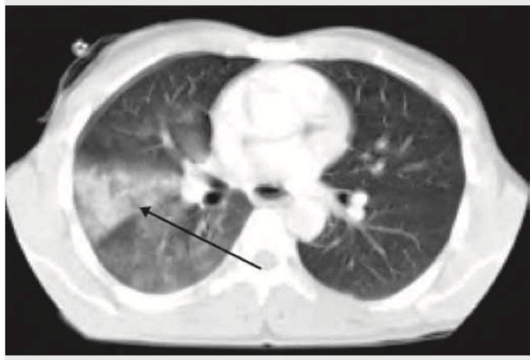
A cement slab fell on the chest of a 20-year-old construction worker. The arrow in the given CT chest points to:
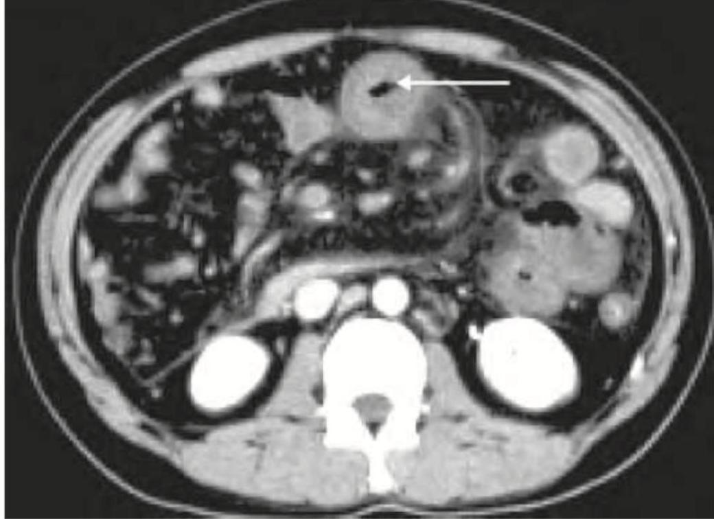
Q50
A 25-year-old patient underwent surgery for scoliosis correction. 5 days post-operatively he develops voluminous bilious vomiting. The given CT abdomen shows: