All (368)Anatomy (30)Anesthesiology (8)Biochemistry (8)Community Medicine (17)Dermatology (24)ENT (18)Forensic Medicine (18)General Medicine (2)Internal Medicine (23)Internal Medicine (8)Microbiology (39)Obstetrics and Gynecology (15)Ophthalmology (16)Orthopaedics (11)Pathology (10)Pathology (17)Pediatrics (26)Pharmacology (6)Physiology (15)Radiology (30)Surgery (5)Surgery (22)
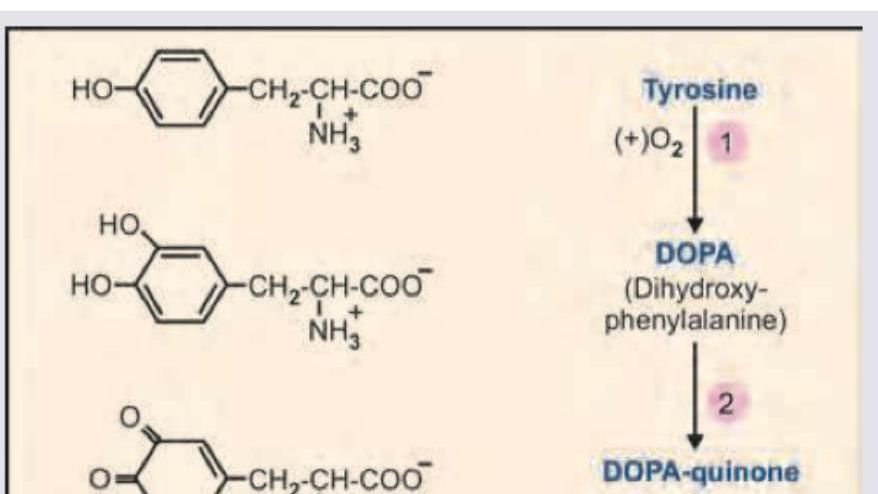
Q331
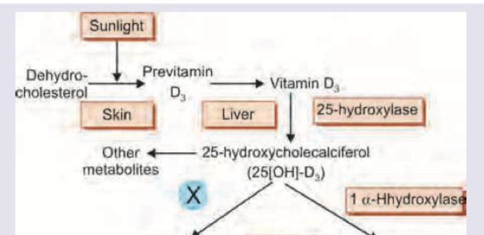
Which of the following is correct about the image shown below?
Q332
Identify the structure marked as X in the specimen of left cubital fossa
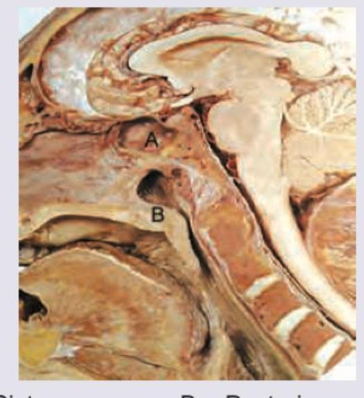
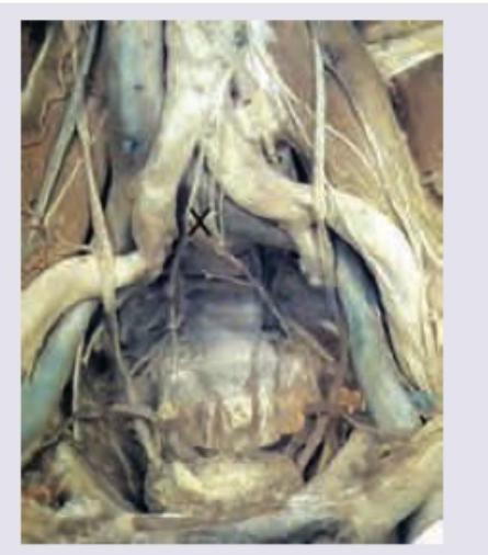
Q333
The following image of posterior abdominal wall and pelvic inlet shows a structure marked as X. Identify it:
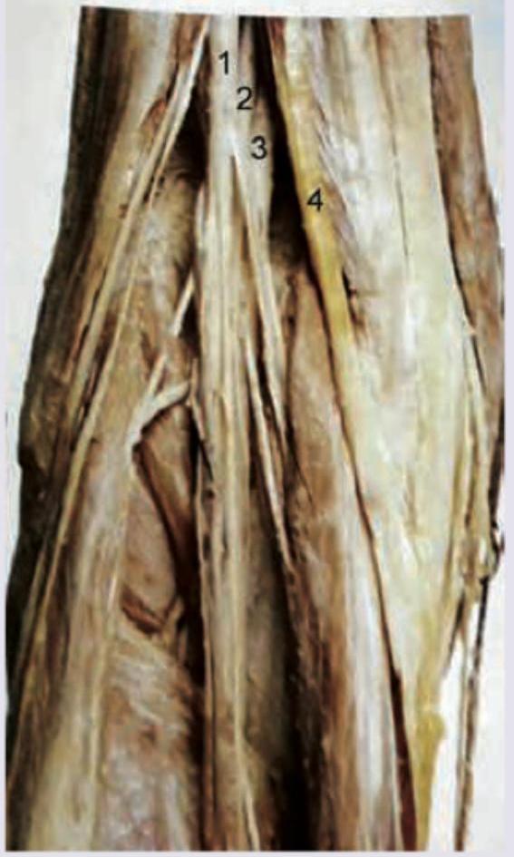
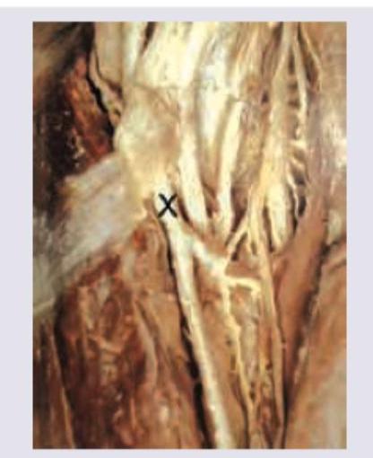
Q334
Which is correct about the markings shown on the left popliteal fossa? (Recent NEET Pattern 2016-17)