All (368)Anatomy (30)Anesthesiology (8)Biochemistry (8)Community Medicine (17)Dermatology (24)ENT (18)Forensic Medicine (18)General Medicine (2)Internal Medicine (23)Internal Medicine (8)Microbiology (39)Obstetrics and Gynecology (15)Ophthalmology (16)Orthopaedics (11)Pathology (10)Pathology (17)Pediatrics (26)Pharmacology (6)Physiology (15)Radiology (30)Surgery (5)Surgery (22)
Q321
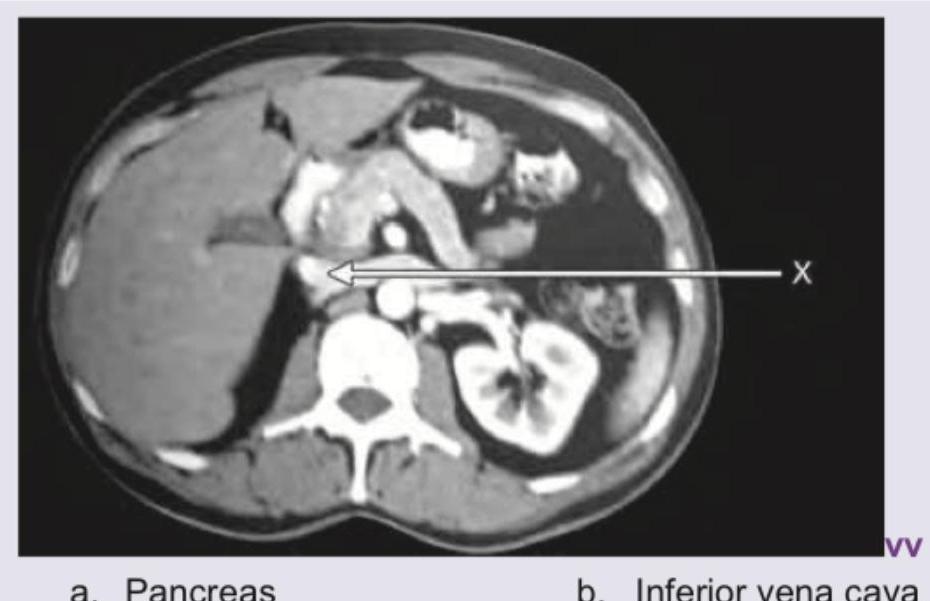
Name the structure marked as $X$ in the CT abdomen shown below: (Recent NEET Pattern 2016-17)
Q322
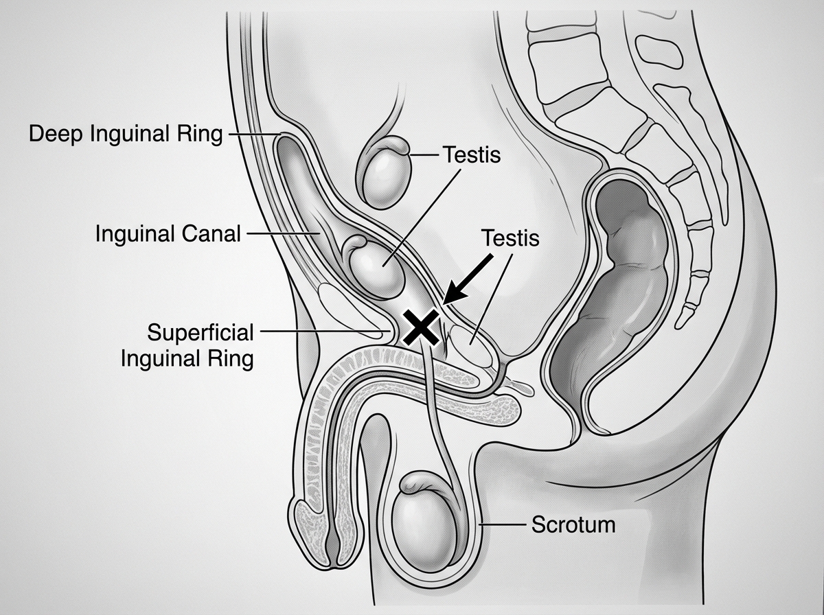
The testis reaches the point marked $X$ at which month of gestation? (Recent NEET Pattern 2016-17)
Q323
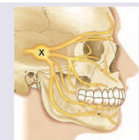
All are true about the ganglion ' $X$ ' shown in the image except:
Q324
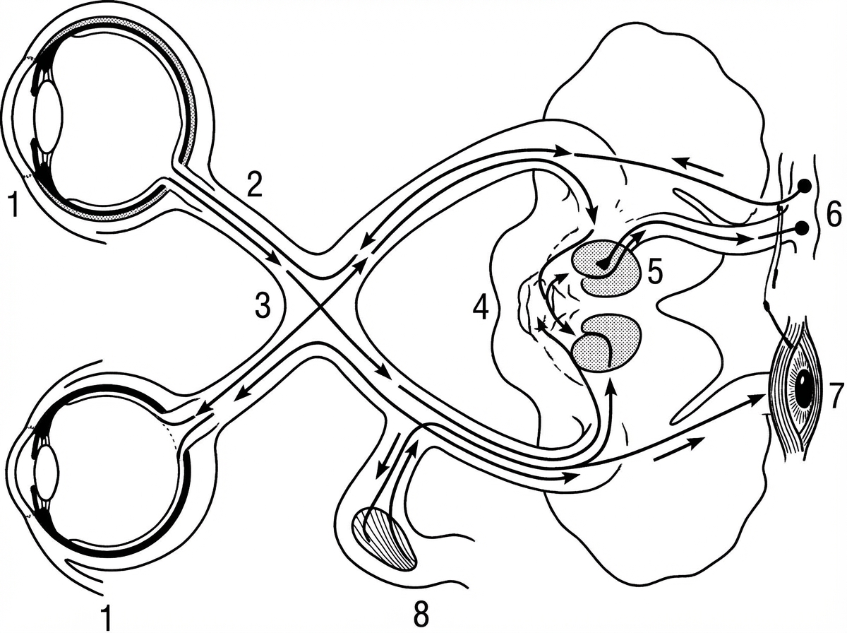
The pathway of light reflex is shown. Lesion of which of the following areas results in development of Argyll Robertson pupil?
Q325
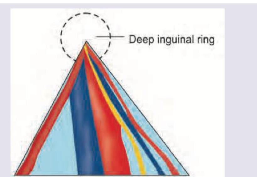
Identify the nerve passing through the Triangle of Doom:
Q326
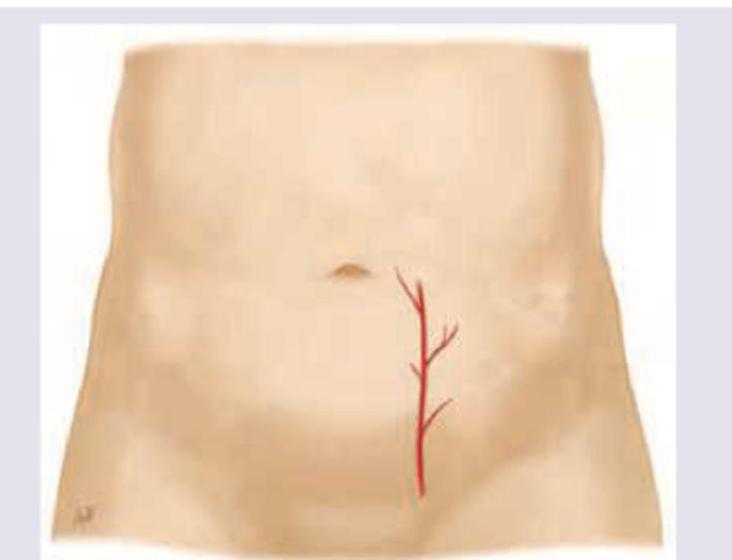
Which artery shown here should be avoided during paracentesis?
Q327
Which is correct sequence about the blood supply of the primitive gut?
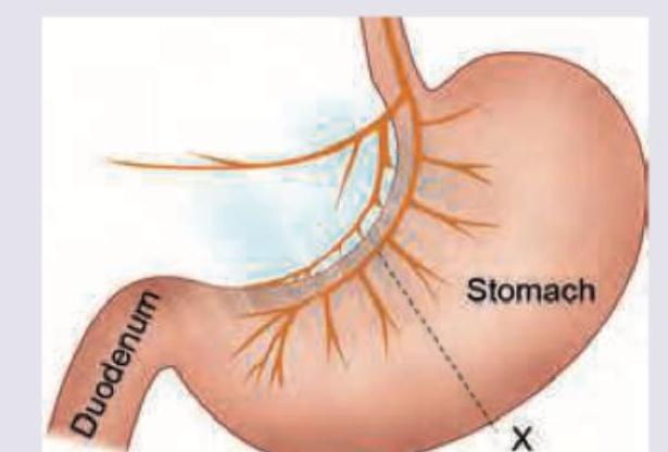
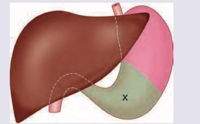
Q328
The area marked as $X$ was selected for gastrostomy. Which of the following statements is incorrect about this area? (Recent NEET Pattern 2016-17)
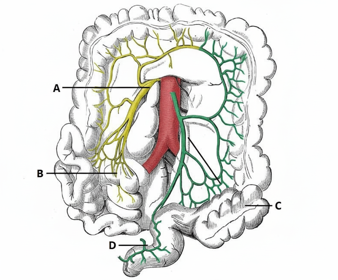
Q329
Which of the following blood vessels is Drummond's Artery? (Recent NEET Pattern 2016-17)