All (368)Anatomy (30)Anesthesiology (8)Biochemistry (8)Community Medicine (17)Dermatology (24)ENT (18)Forensic Medicine (18)General Medicine (2)Internal Medicine (23)Internal Medicine (8)Microbiology (39)Obstetrics and Gynecology (15)Ophthalmology (16)Orthopaedics (11)Pathology (10)Pathology (17)Pediatrics (26)Pharmacology (6)Physiology (15)Radiology (30)Surgery (5)Surgery (22)
Q311
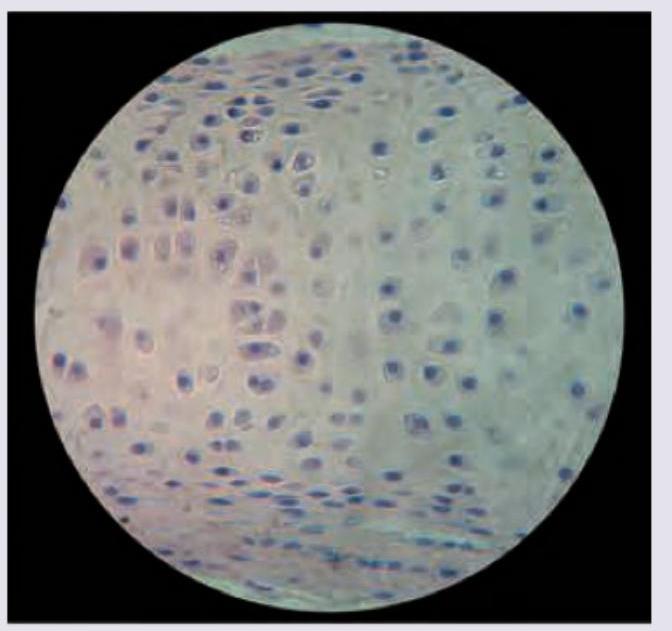
All are true about the cartilage shown in the figure except: (Recent NEET Pattern 2016-17)
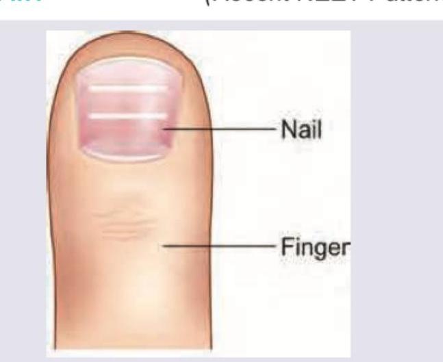
Q312
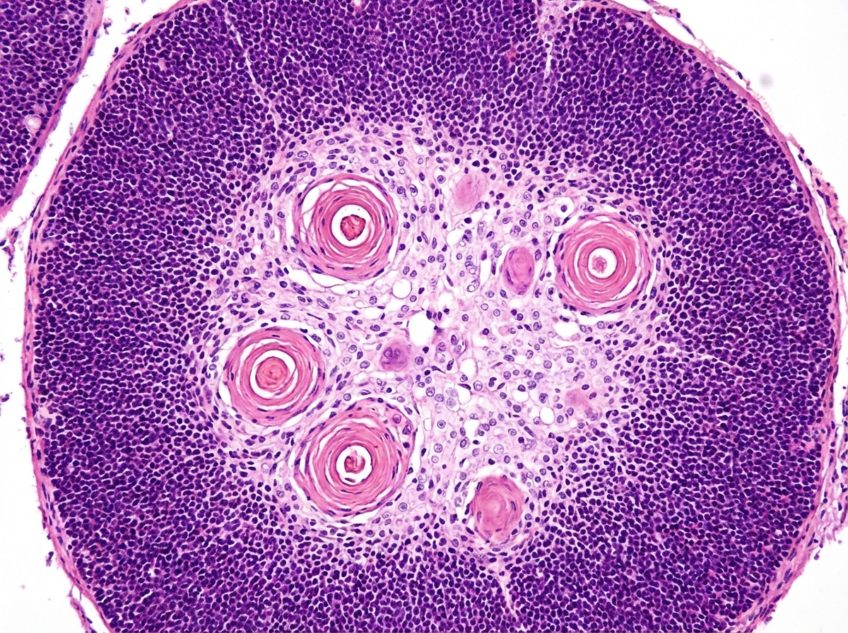
Identify the structure:
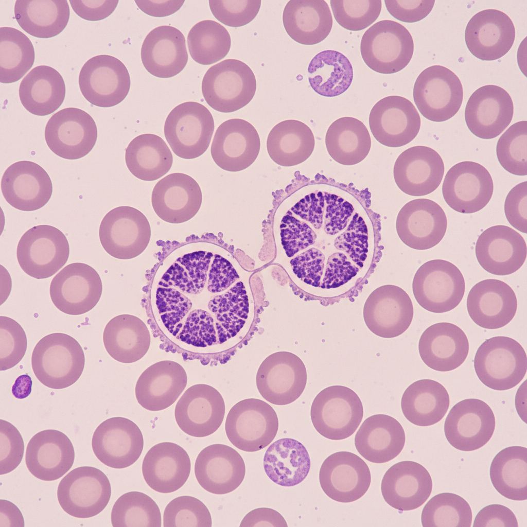
Q313
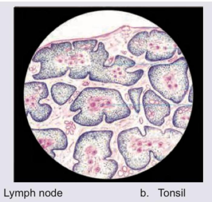
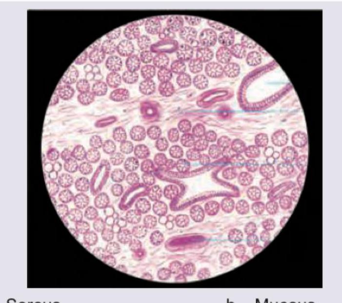
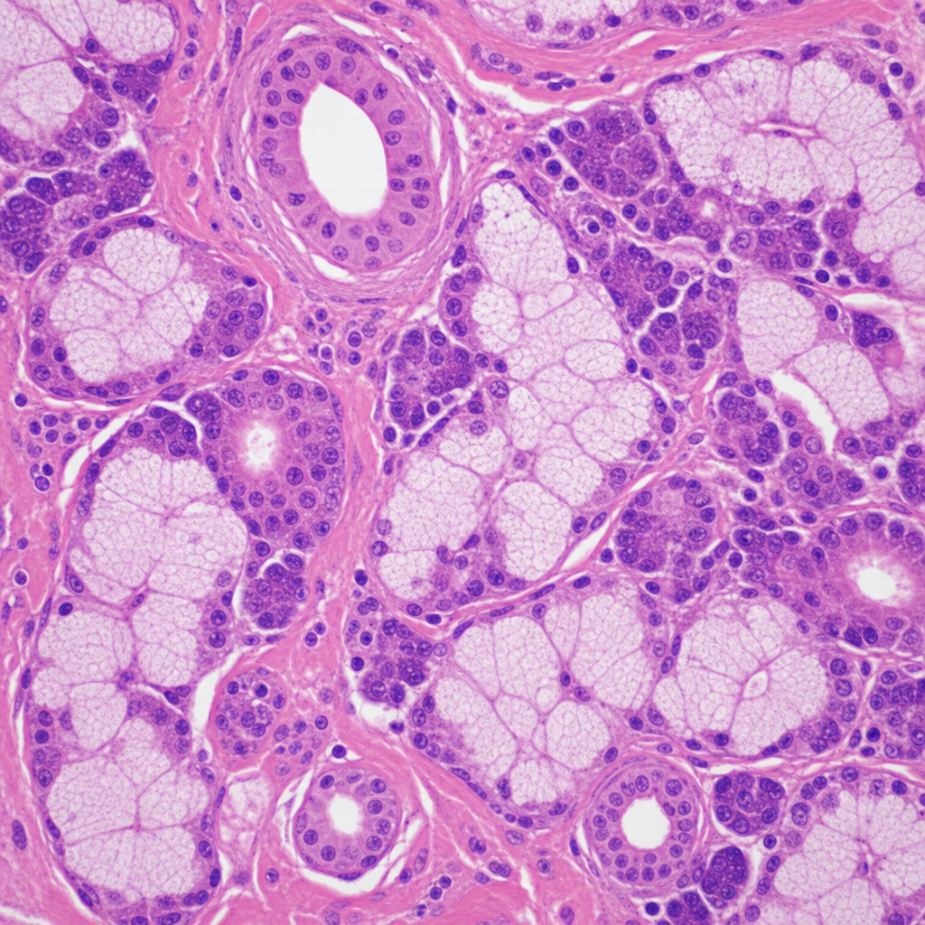
Which type of salivary glands is shown in the image?
Q314
Which type of salivary gland is shown in the image?
Q315
Which type of salivary glands is shown in the image?
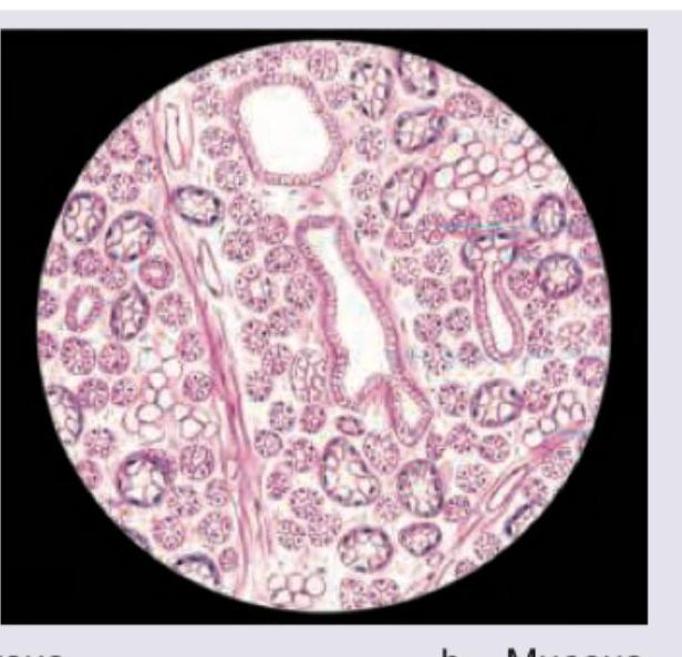
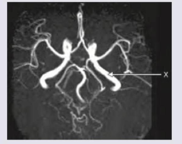
Q316
The blood vessel marked as $X$ in the CT angiography image is: