All (368)Anatomy (30)Anesthesiology (8)Biochemistry (8)Community Medicine (17)Dermatology (24)ENT (18)Forensic Medicine (18)General Medicine (2)Internal Medicine (23)Internal Medicine (8)Microbiology (39)Obstetrics and Gynecology (15)Ophthalmology (16)Orthopaedics (11)Pathology (10)Pathology (17)Pediatrics (26)Pharmacology (6)Physiology (15)Radiology (30)Surgery (5)Surgery (22)
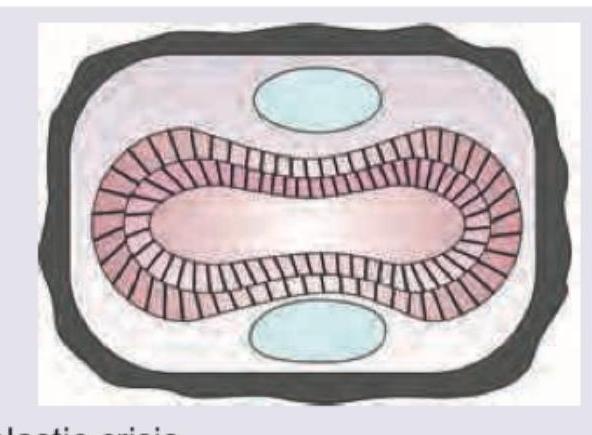
Q241
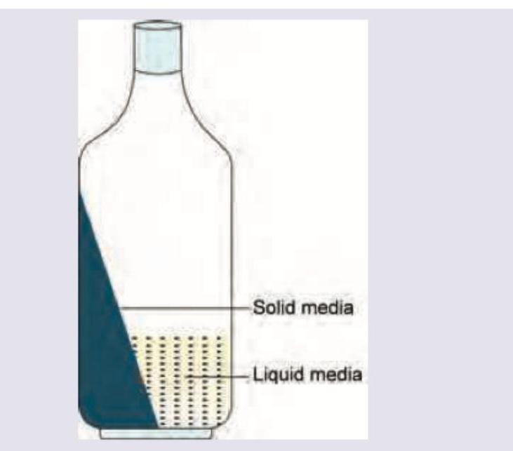
The following two phase culture system is used for diagnosis of: (Recent NEET Pattern 2016-17)
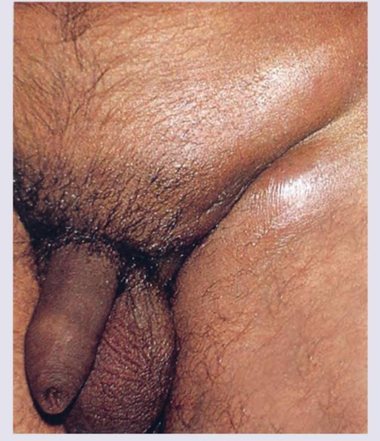
Q242
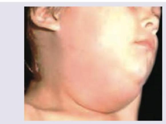
All are correct about the condition shown in the image except:
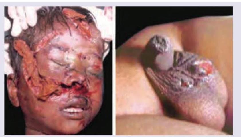
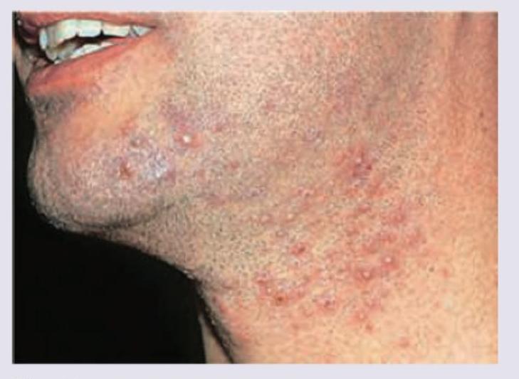
Q243
Which organism is incriminated in causing the following lesions? (Recent NEET Pattern 2016-17)
Q244
Which of the following organisms is incriminated in a patient of left sided endocarditis involving the mitral valve? (Recent NEET Pattern 2016-17)
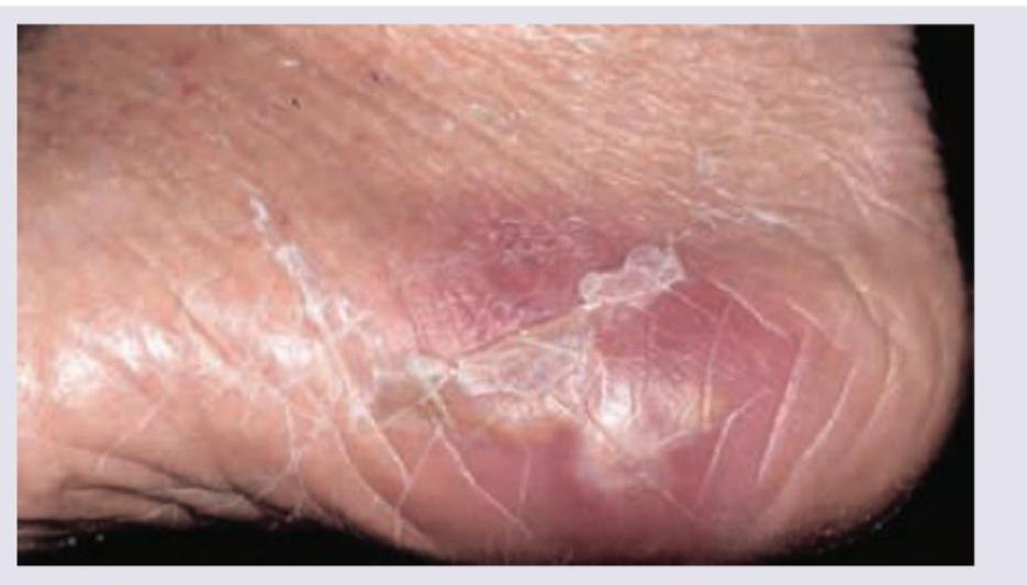
Q245
A patient walking barefoot during his morning walk has developed a swelling in the foot. What is the probable diagnosis?
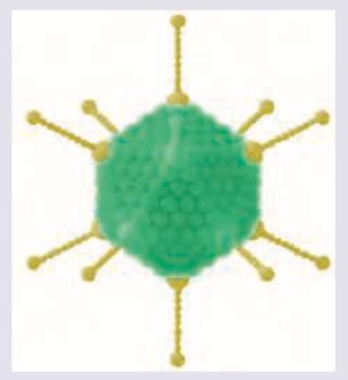
Q246
Which of the following diseases is caused by the virus shown below?
Q247
Which of the following diseases is caused by the virus shown below?
Q248
A 2-year-old unimmunized child from a village presents with fever, decreased feeding and ear ache. All are true about the virus responsible for the condition shown except: (Recent NEET Pattern 2016-17)
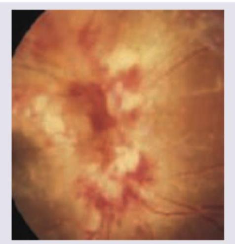
Q249
A 40-year-old AIDS positive patient complains of seeing floaters followed by progressive reduction in visual acuity over next weeks. Fundus examination was performed. All are true about the causative agent except: (Recent NEET Pattern 2016-17)