All (366)Anatomy (30)Anesthesiology (8)Biochemistry (8)Community Medicine (16)Dermatology (24)ENT (18)Forensic Medicine (18)General Medicine (2)Internal Medicine (23)Internal Medicine (8)Microbiology (39)Obstetrics and Gynecology (15)Ophthalmology (16)Orthopaedics (10)Pathology (10)Pathology (17)Pediatrics (26)Pharmacology (6)Physiology (15)Radiology (30)Surgery (5)Surgery (22)
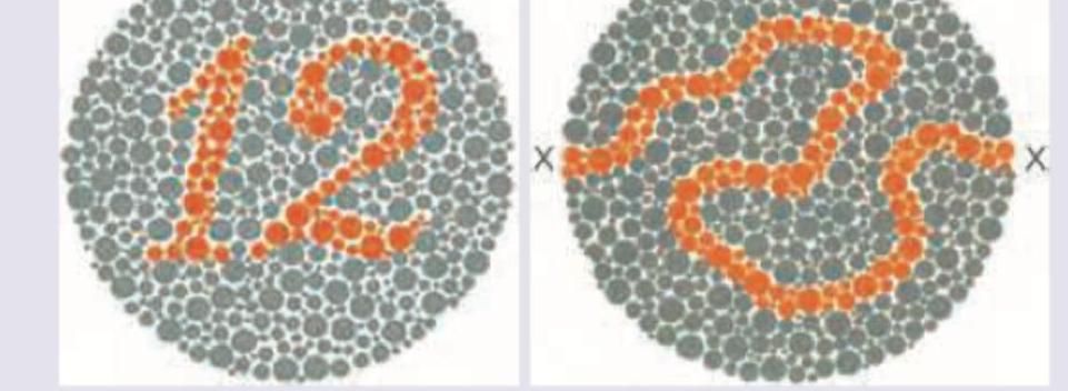
Q181
The chart shown in the image is:
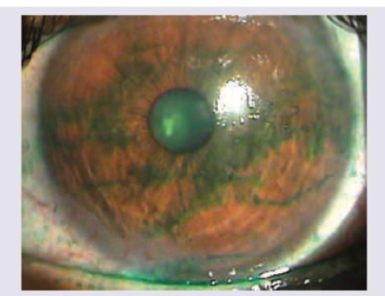
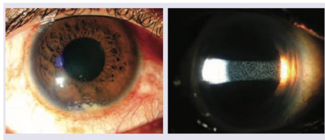
Q182
Identify the stain instilled in the eye in the following image:
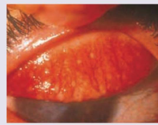
Q183
A patient from slum presents with grittiness in eyes. Everted eyelid shows: (Recent NEET Pattern 2016-17)
Q184
All are causes of the presentation shown below except:
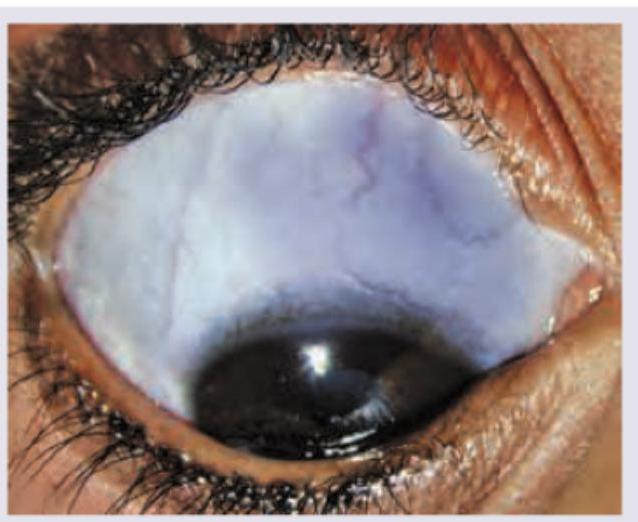
Q185
A 30-year-old school teacher presents with complaints of red eye with photophobia. Ocular findings are shown below. All are true about the condition shown except: (Recent NEET Pattern 2016-17)
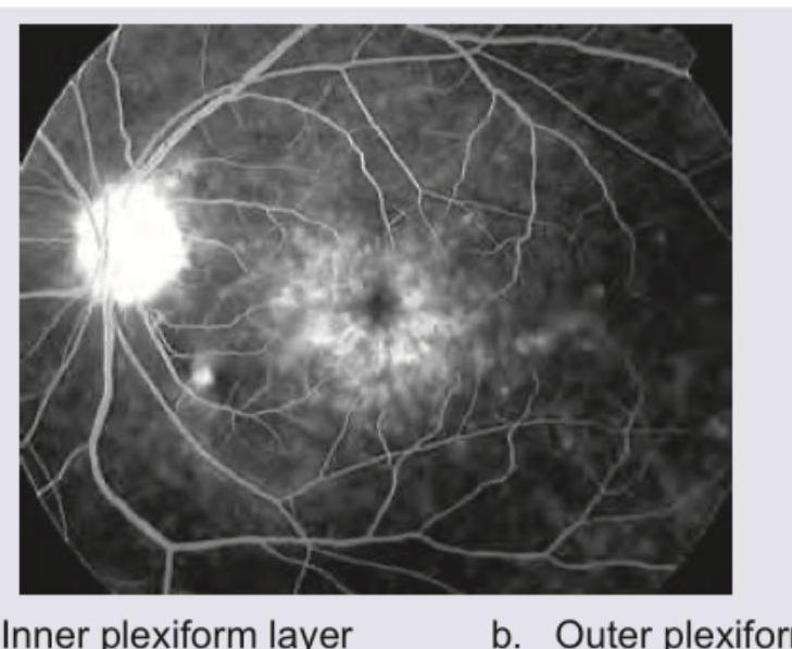
Q186
The given FFA appearance occurs due to accumulation of dye in which of the following layers of retina?
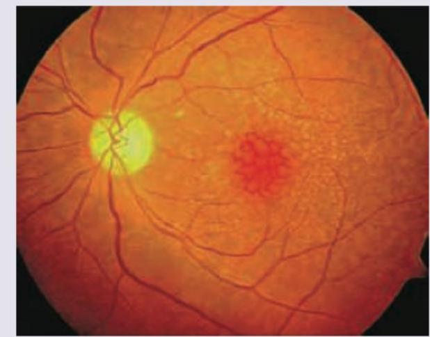
Q187
A 75-year-old Englishman living in India presents to OPD with complaints of gradual onset painless, progressive blurring of central vision. He says he could earlier drive to the hospital by himself but is not able to do so now. Slit lamp examination is normal. Fundus examination is given below. What is the diagnosis?
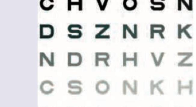
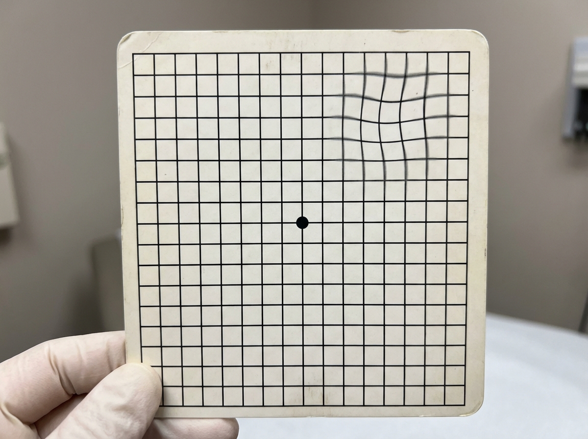
Q188
The test shown below is used for the evaluation of
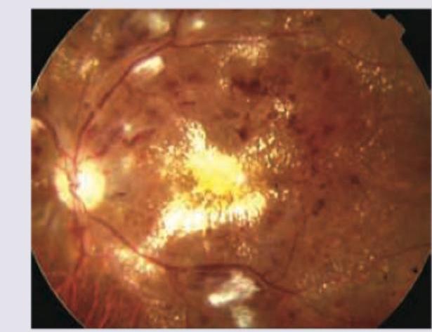
Q189
A 60-year-old patient during annual check-up had a report of HbA1C of $10 \%$. What does the given fundus examination show?