All SubjectsAnatomy (30)Anesthesiology (8)Biochemistry (8)Community Medicine (16)Dermatology (24)ENT (18)Forensic Medicine (18)General Medicine (2)Internal Medicine (23)Internal Medicine (8)Microbiology (39)Obstetrics and Gynecology (15)Ophthalmology (16)Orthopaedics (11)Pathology (10)Pathology (17)Pediatrics (26)Pharmacology (6)Physiology (15)Radiology (30)Surgery (5)Surgery (22)
Q11
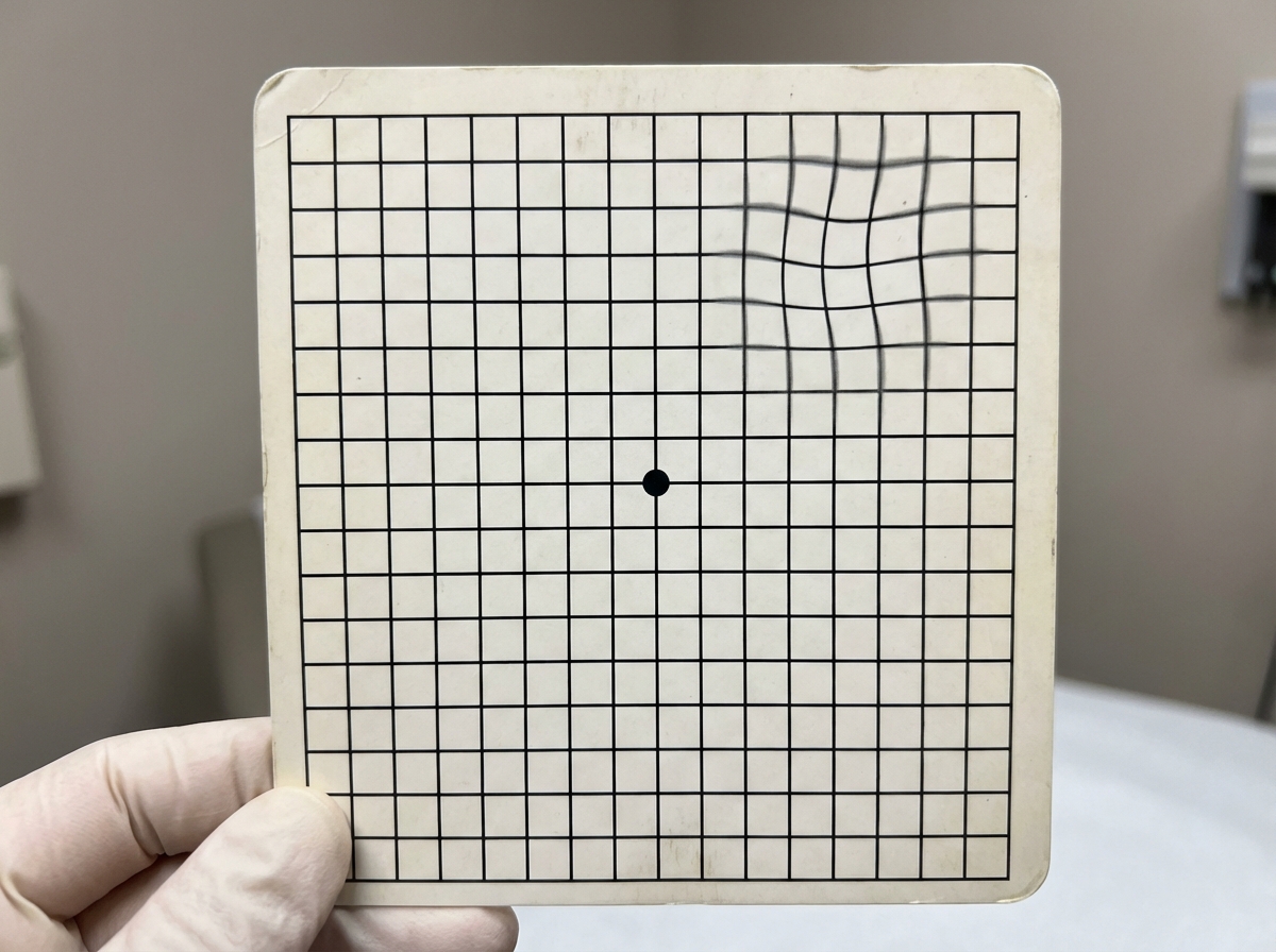
The test shown below is used for the evaluation of
Q12
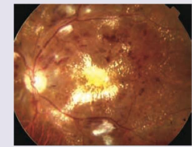
A 60-year-old patient during annual check-up had a report of HbA1C of $10 \%$. What does the given fundus examination show?
Q13
All are used for the treatment of the condition shown below except:
Q14
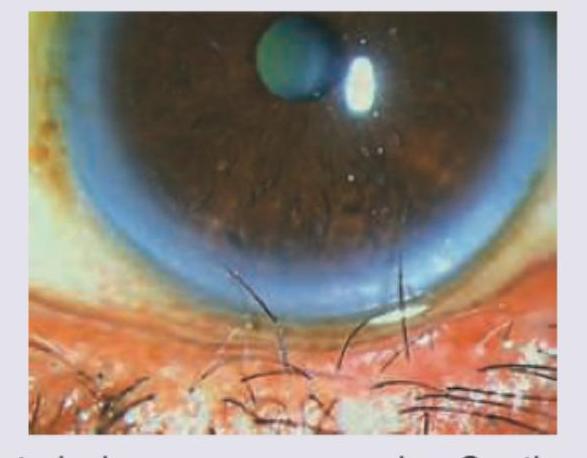
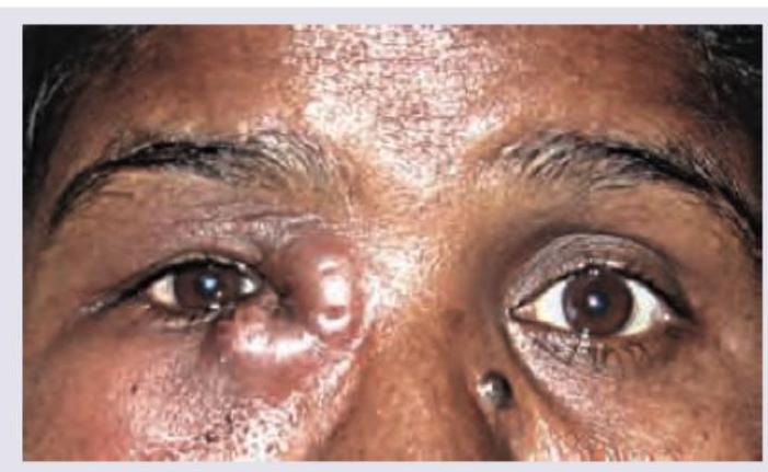
A 30-year-old woman presents with painful eye swelling. Based on the clinical photograph shown, the most likely diagnosis is:
Q15
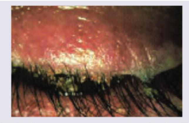
A patient presents with itching in eyes with redness of eyelids. What is correct about the image shown below? (Recent NEET Pattern 2016-17)
Q16
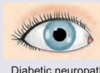
A 20-year-old woman is admitted with the following presentation. 1% pilocarpine is not showing any response on the side of mydriasis. What is the diagnosis? (Recent NEET Pattern 2016-17)