All SubjectsAnatomy (30)Anesthesiology (8)Biochemistry (8)Community Medicine (17)Dermatology (24)ENT (18)Forensic Medicine (18)General Medicine (2)Internal Medicine (23)Internal Medicine (8)Microbiology (39)Obstetrics and Gynecology (15)Ophthalmology (16)Orthopaedics (11)Pathology (10)Pathology (17)Pediatrics (26)Pharmacology (6)Physiology (15)Radiology (30)Surgery (5)Surgery (22)
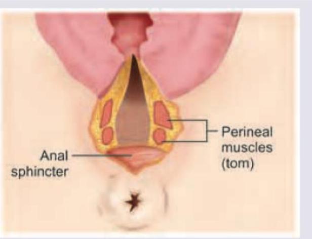
Q11
Which degree of obstetric anal sphincter injury is seen here?
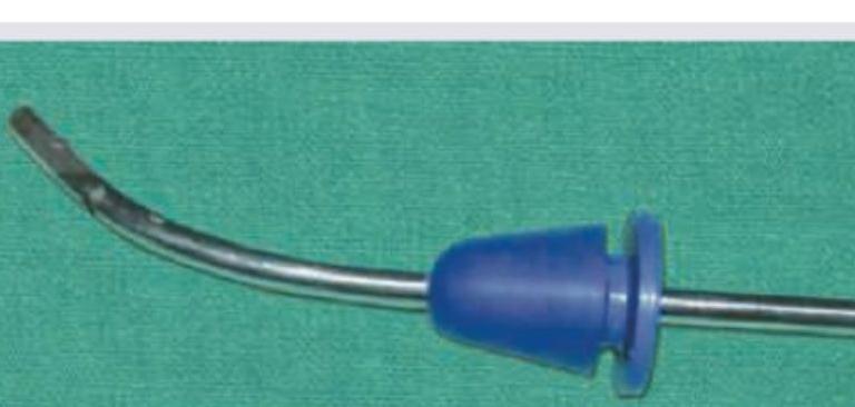
Q12
All are correct regarding the device shown here except:
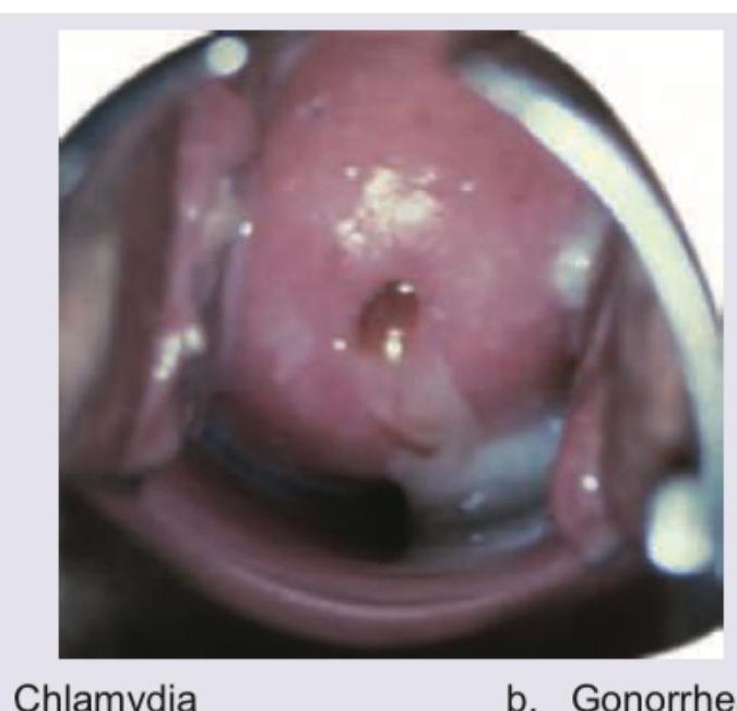
Q13
Identify the STD. (Recent Neet Pattern 2016-17)
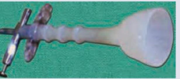
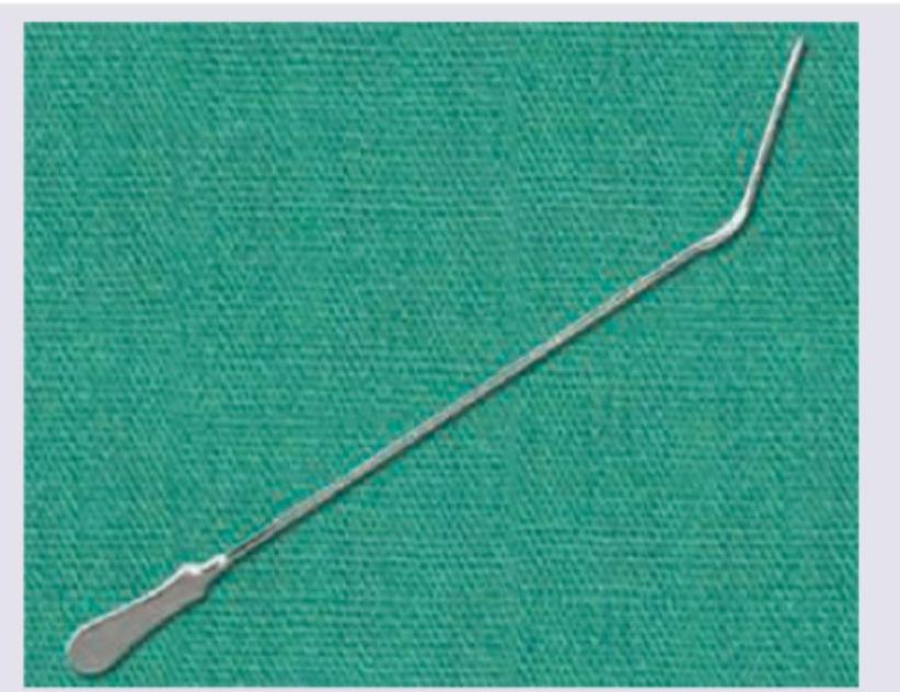
Q14
Which of the following is incorrect about the instrument shown below? (Recent Neet Pattern 2016-17)
Q15
Identify the instrument: