All are correct about the organism causing the following lesion except:
Which of the following is correct about the vegetative fungal spores?
The image shows:
Which of the following is correct about the media shown below?
The following diagram depicts blood smear of which species?
NEET-PG 2017 - Microbiology NEET-PG Practice Questions and MCQs
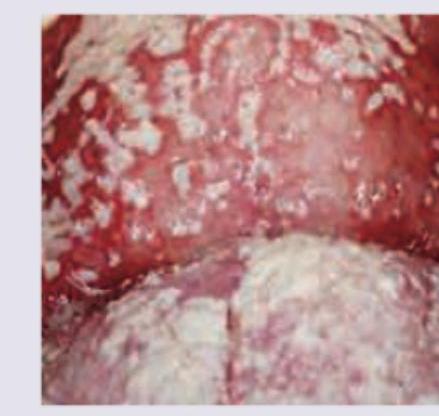
Question 21: All are correct about the organism causing the following lesion except:
- A. Reynold Braude phenomenon (Correct Answer)
- B. Gram positive yeast cells
- C. Chlamydospores obtained on cornmeal agar
- D. Creamy patches that on removal lead to red oozing patches
Explanation: ***Reynold Braude phenomenon*** - The **Reynolds-Braude phenomenon** is associated with **Staphylococcus aureus** and refers to the increased invasiveness of *S. aureus* in the presence of certain other bacteria. - This phenomenon is **not characteristic of *Candida albicans***, the organism responsible for the oral candidiasis (thrush) shown in the image. - **This is the correct answer** to this "except" question. *Gram positive yeast cells* - *Candida albicans* is a **Gram-positive yeast** that appears purple/blue on Gram staining. - It typically shows **budding yeast cells** and **pseudohyphae** on microscopy. - This statement is TRUE for *Candida albicans*. *Chlamydospores obtained on cornmeal agar* - **Chlamydospores** are thick-walled, large, round terminal spores that are characteristic of *Candida albicans*. - They are best demonstrated on **cornmeal agar with Tween 80** (or rice agar), which is the standard medium for their production. - The presence of chlamydospores is a key identifying feature that helps differentiate *C. albicans* from other *Candida* species. - This statement is TRUE for *Candida albicans*. *Creamy patches that on removal lead to red oozing patches* - The image shows **creamy white patches** on an inflamed oral mucosa, characteristic of **pseudomembranous candidiasis** (oral thrush). - These patches can be **scraped off**, revealing an **erythematous, bleeding** (red oozing) underlying surface. - This is a classic clinical presentation of oral candidiasis. - This statement is TRUE for *Candida albicans*.
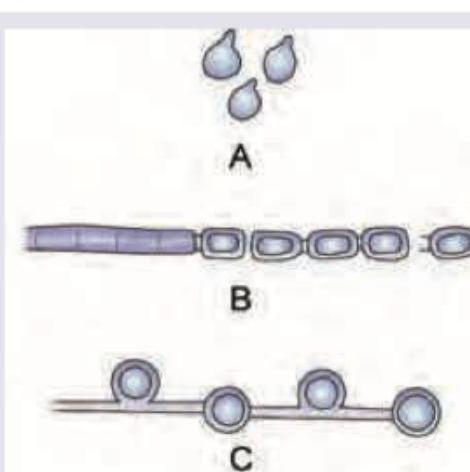
Question 22: Which of the following is correct about the vegetative fungal spores?
- A. A = Arthrospores, B= Blastospores, C= Chlamydospores
- B. A = Blastospores, B= Arthrospores, C= Chlamydospores (Correct Answer)
- C. A = Blastospores, B= Chlamydospores, C= Arthrospores
- D. A = Chlamydospores, B= Arthrospores, C= Blastospores
Explanation: **A = Blastospores, B= Arthrospores, C= Chlamydospores** - Image A depicts **blastospores**, which are asexually produced spores formed by **budding** from a parent cell, giving them a distinct tear-drop or oval shape. - Image B illustrates **arthrospores**, which are formed by the **fragmentation** of a hyphal cell into barrel-shaped segments. - Image C shows **chlamydospores**, characterized by their **thick-walled**, resistant, and usually spherical or oval structure within a hypha. *A = Arthrospores, B= Blastospores, C= Chlamydospores* - This option incorrectly identifies image A as arthrospores, which are typically barrel-shaped and result from hyphal fragmentation, not the budding pattern seen in image A. - Image B is incorrectly labeled as blastospores, but the fragmentation pattern is characteristic of arthrospores. *A = Blastospores, B= Chlamydospores, C= Arthrospores* - While image A is correctly identified as blastospores, this option misidentifies image B as chlamydospores. - Image C does not show arthrospores; the thick-walled structure is characteristic of chlamydospores, not the barrel-shaped arthrospores. *A = Chlamydospores, B= Arthrospores, C= Blastospores* - This option incorrectly identifies image A as chlamydospores, which are thick-walled resistant structures, not the budding spores visible in the image. - It also incorrectly labels image C as blastospores; the thick-walled appearance is typical of chlamydospores, not budded blastospores.
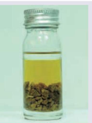
Question 23: The image shows:
- A. Alkaline peptone water
- B. Lowenstein Jensen media
- C. Cary Blair medium
- D. Robertson cooked meat broth (Correct Answer)
Explanation: ***Robertson cooked meat broth*** - The image displays a glass jar containing a **meat-based medium** at the bottom, covered with a liquid, which is characteristic of Robertson cooked meat broth. - This medium is primarily used for the **cultivation of anaerobic bacteria**, as the meat particles create an anaerobic environment and provide nutrients. *Alkaline peptone water* - Alkaline peptone water is a **liquid enrichment medium** that appears as a clear, yellowish broth without any particulate matter like cooked meat. - It is specifically used for the enrichment of *Vibrio cholerae* due to its high pH. *Lowenstein Jensen media* - Lowenstein Jensen media is a **solid egg-based medium** that is typically green in color and typically slants in test tubes or petri dishes. - It is used for the isolation and culture of **mycobacteria**, and its appearance is distinctly different from the image. *Cary Blair medium* - Cary Blair medium is a **semisolid transport medium** used for the preservation of enteric bacteria in stool samples. - It appears as a clear or slightly opalescent gel in a tube and does not contain visible chunks of meat.
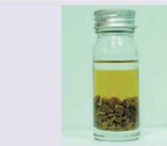
Question 24: Which of the following is correct about the media shown below?
- A. Saccharolytic bacteria turn meat black
- B. Proteolytic bacteria turn meat red
- C. Liquid paraffin layering over the broth prevents air entry (Correct Answer)
- D. Favor growth of microaerophilic bacteria
Explanation: ***Liquid paraffin layering over the broth prevents air entry*** - The image depicts **Cooked Meat Medium (CMM)**, which is used primarily for cultivating **anaerobic bacteria**, particularly Clostridia species. - **Liquid paraffin layering** on top of the broth is a key feature of CMM that **creates anaerobic conditions** by preventing oxygen diffusion into the medium. - This technique, combined with the reducing substances from meat particles, ensures a truly anaerobic environment suitable for obligate anaerobes. *Saccharolytic bacteria turn meat black* - **Saccharolytic bacteria** primarily ferment carbohydrates and produce acid/gas, but do not typically cause blackening of meat. - Blackening in CMM is associated with **hydrogen sulfide (H2S) production** by proteolytic bacteria, not saccharolytic activity. *Proteolytic bacteria turn meat red* - This is incorrect. **Proteolytic bacteria** break down proteins and typically turn the meat particles **black** due to H2S production. - The digestion of meat proteins by proteolytic Clostridia results in darkening (blackening), not reddening of the medium. *Favor growth of microaerophilic bacteria* - CMM is designed for **anaerobic bacteria**, not microaerophiles. - **Microaerophilic bacteria** require low oxygen tension (5-10% O2) and are cultivated in specialized conditions like candle jars or microaerophilic atmosphere generators, not in CMM. - CMM provides an anaerobic environment through liquid paraffin seal and reducing agents from meat.
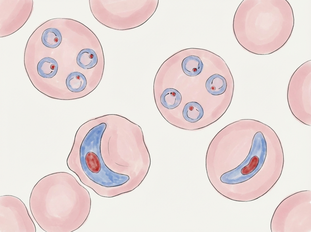
Question 25: The following diagram depicts blood smear of which species?
- A. Plasmodium malariae
- B. Plasmodium ovale
- C. Plasmodium falciparum (Correct Answer)
- D. Plasmodium vivax
Explanation: ***Plasmodium falciparum*** - The image displays characteristic **crescent-shaped or banana-shaped gametocytes** (stages 16-20), which are **pathognomonic for *Plasmodium falciparum*** - The earlier stages (1-5) represent ring forms (multiple rings per RBC, delicate ring forms) - Subsequent stages (6-15) show maturation culminating in the distinct **crescentic gametocytes** - This morphology is the **key distinguishing feature** of P. falciparum *Plasmodium vivax* - Produces **oval-shaped gametocytes**, not crescent-shaped - Characterized by enlarged RBCs with **Schüffner's dots** - Shows amoeboid trophozoites, not seen in this smear *Plasmodium malariae* - Produces **oval to round gametocytes** - Characteristic **band forms** and **rosette-shaped schizonts** (8-12 merozoites) - Not crescent-shaped as shown in the image *Plasmodium ovale* - Produces **oval gametocytes** similar to P. vivax - RBCs show **fimbriated edges** and Schüffner's dots - Does not produce crescent-shaped gametocytes