All SubjectsAnatomy (30)Anesthesiology (8)Biochemistry (8)Community Medicine (17)Dermatology (24)ENT (18)Forensic Medicine (18)General Medicine (2)Internal Medicine (23)Internal Medicine (8)Microbiology (39)Obstetrics and Gynecology (15)Ophthalmology (16)Orthopaedics (11)Pathology (10)Pathology (17)Pediatrics (26)Pharmacology (6)Physiology (15)Radiology (30)Surgery (5)Surgery (22)
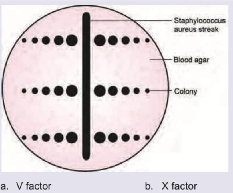
Q11
The following plate shows *H. influenzae* colonies. Which of the following is responsible for the phenomenon shown? (Recent NEET Pattern 2016-17)
Q12
All are true about the organism whose colonies are shown below except:
Q13
A 35-year-old patient presents to the OPD 24 hours after a fight with a stranger in which he was bitten. GCS is 15/15 and following injury is noted on left forearm. He complains of extreme pain and tenderness in the injury. Swab from the injury was plated in chocolate agar and incubated in 10% carbon dioxide for 48 hours. Small colonies with pitting appearance were noted. Which of the following organism is responsible?
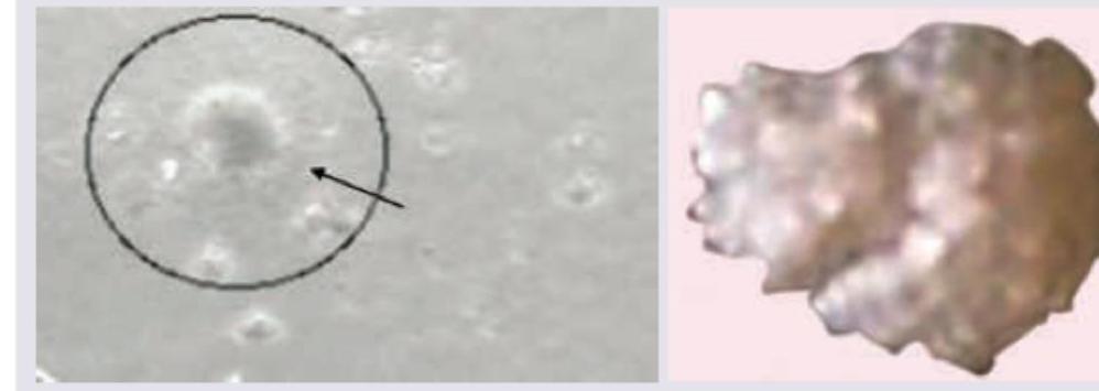
Q14
The following organism shown leads to development of:
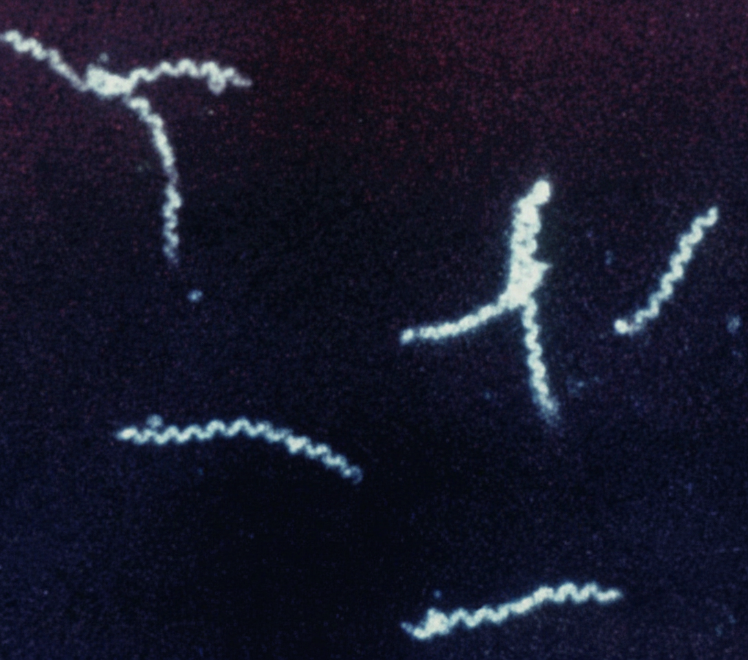
Q15
All are correct about the organism shown in the image except: (Recent NEET Pattern 2016-17)
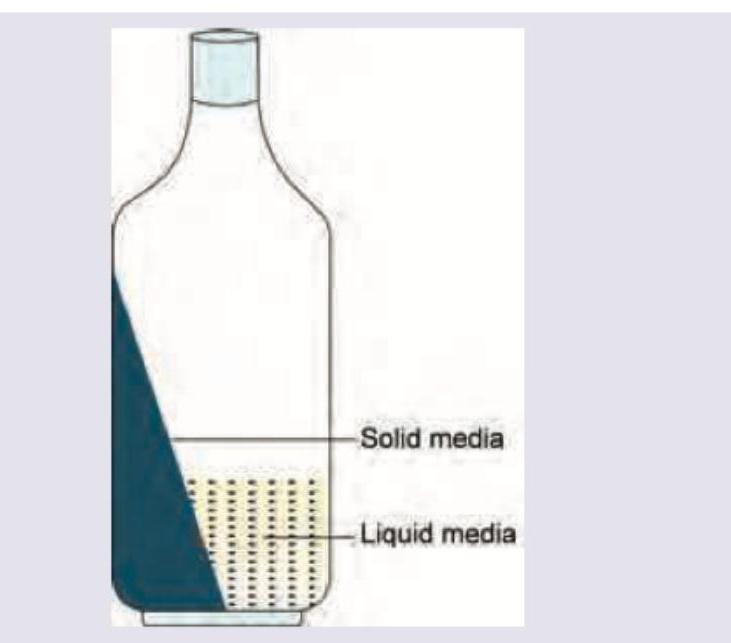
Q16
The following two phase culture system is used for diagnosis of: (Recent NEET Pattern 2016-17)
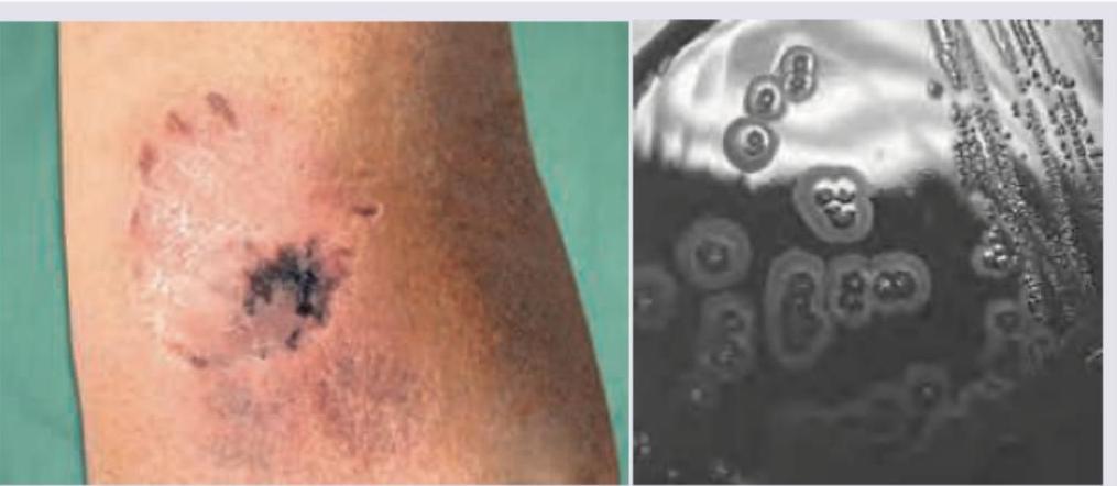
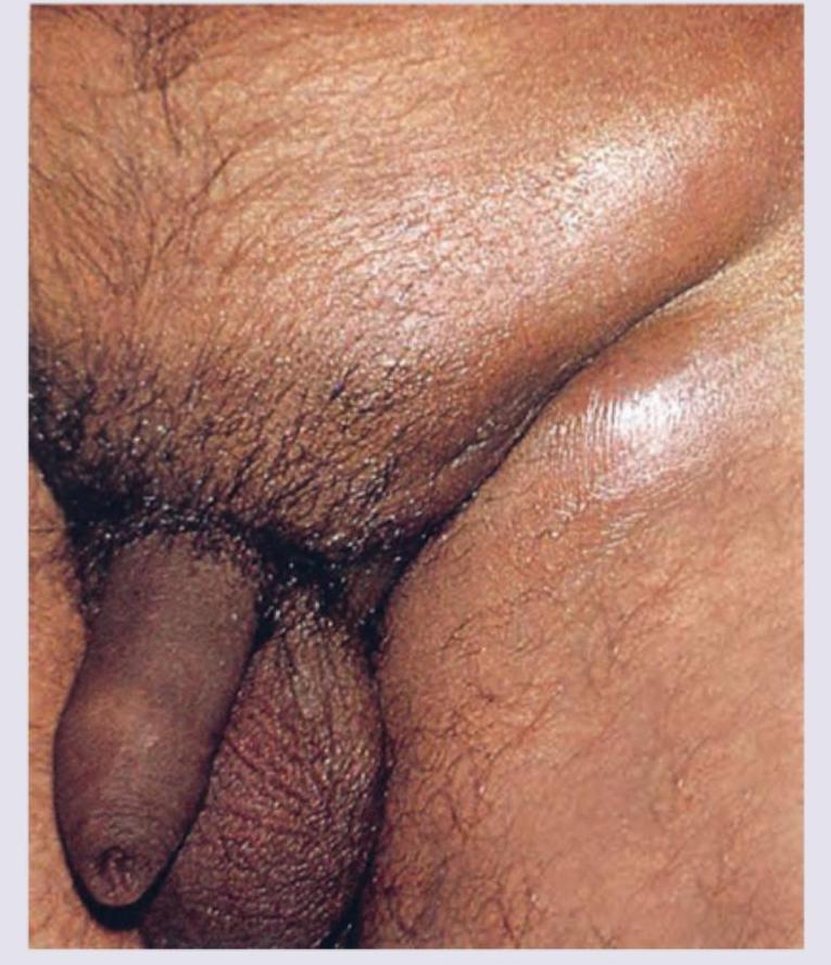
Q17
All are correct about the condition shown in the image except:
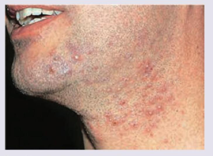
Q18
Which organism is incriminated in causing the following lesions? (Recent NEET Pattern 2016-17)
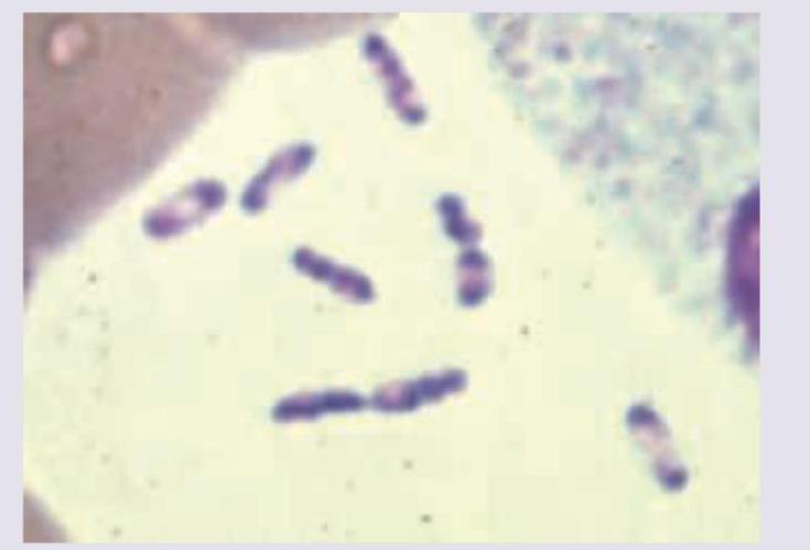
Q19
Which of the following organisms is incriminated in a patient of left sided endocarditis involving the mitral valve? (Recent NEET Pattern 2016-17)
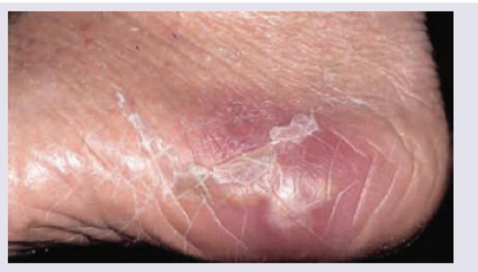
Q20
A patient walking barefoot during his morning walk has developed a swelling in the foot. What is the probable diagnosis?