NEET-PG 2017 — Microbiology
25 Previous Year Questions with Answers & Explanations
All are true about Helicobacter pylori on special stain preparation of stomach except:
All are true about bacteria shown in the smear of pus below, except:
All are true about the bacteria shown in the figure except:
All of the following are caused by the organism shown except:
What is the drug of choice for organism producing the following colonies?
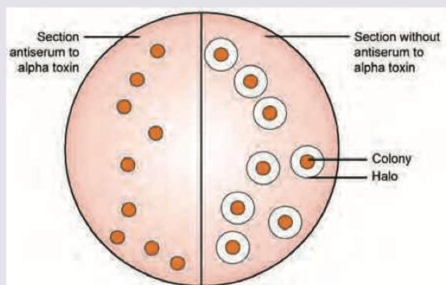
What is the following pattern seen with *Clostridium perfringens* called?
Which of the bacteria is shown in this culture plate of nutrient agar? (Recent NEET Pattern 2016-17)
The following plate shows *H. influenzae* colonies. Which of the following is responsible for the phenomenon shown? (Recent NEET Pattern 2016-17)
All are true about the organism whose colonies are shown below except:
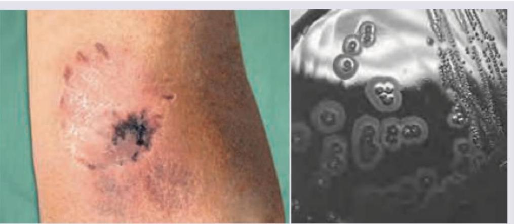
A 35-year-old patient presents to the OPD 24 hours after a fight with a stranger in which he was bitten. GCS is 15/15 and following injury is noted on left forearm. He complains of extreme pain and tenderness in the injury. Swab from the injury was plated in chocolate agar and incubated in 10% carbon dioxide for 48 hours. Small colonies with pitting appearance were noted. Which of the following organism is responsible?
NEET-PG 2017 - Microbiology NEET-PG Practice Questions and MCQs
Question 1: All are true about Helicobacter pylori on special stain preparation of stomach except:
- A. Steiner stain preparation
- B. S shaped non-flagellated bacteria (Correct Answer)
- C. Dormant stage is coccoid form
- D. Attaches but does not invade the cells
Explanation: ***S shaped non-flagellated bacteria*** - *H. pylori* are generally **spiral-shaped** or **curved rods**, not typically S-shaped, and are characterized by their **polar flagella** which are essential for their motility in the viscous gastric mucus. - The presence of flagella is a key feature distinguishing *H. pylori* and enabling its survival in the stomach environment. *Steiner stain preparation* - The **Steiner silver stain** is commonly used to visualize *H. pylori* in gastric biopsies, demonstrating them as dark, helical organisms. - While effective, other stains like Giemsa or Warthin-Starry are also used, but Steiner stain is a valid method for detection. *Dormant stage is coccoid form* - Under stressful conditions, such as antibiotic exposure or prolonged culture, *H. pylori* can transform into a **coccoid form**. - This coccoid form is considered a **viable but non-culturable** dormant stage, potentially contributing to persistence and recurrence of infection. *Attaches but does not invade the cells* - *H. pylori* colonizes the **mucus layer** and attaches to the apical surface of gastric epithelial cells but generally **does not invade** the cells. - Its effects are mediated by toxins and enzymes released into the extracellular space, leading to inflammation and cellular damage without direct intracellular invasion.
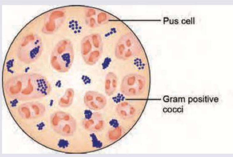
Question 2: All are true about bacteria shown in the smear of pus below, except:
- A. Produce black colonies on potassium tellurite blood agar
- B. Most strains are inhibited in presence of 5 % NaCl (Correct Answer)
- C. Liquefy gelatin
- D. Seen in Job syndrome
Explanation: The image shows **Gram-positive cocci** arranged in clusters, which is characteristic of *Staphylococcus aureus*. Many pus cells (neutrophils) are also seen, indicating an inflammatory response, consistent with a bacterial infection. ***Most strains are inhibited in presence of 5 % NaCl*** - *Staphylococcus aureus* is known to be **halophilic**, meaning it **grows well** in the presence of high salt concentrations (e.g., 7.5% NaCl), which distinguishes it from many other bacteria. - Therefore, the statement that it is 'inhibited in the presence of 5% NaCl' is **incorrect**. *Produce black colonies on potassium tellurite blood agar* - *Staphylococcus aureus* **reduces tellurite** to metallic tellurium, resulting in the production of **black colonies** on potassium tellurite blood agar. - This is a characteristic feature used in the identification of *Staphylococcus aureus*, particularly on media like Baird-Parker agar. *Liquefy gelatin* - *Staphylococcus aureus* produces the enzyme **gelatinase**, which **hydrolyzes gelatin**, causing it to liquefy. - This is a biochemical characteristic used to differentiate *Staphylococcus aureus* from certain other staphylococcal species. *Seen in Job syndrome* - **Job syndrome** (Hyper-IgE syndrome) is characterized by recurrent severe **Staphylococcal infections** of the skin and lungs. - Patients with Job syndrome have impaired neutrophil chemotaxis and elevated IgE levels, making them highly susceptible to *Staphylococcus aureus* infections.
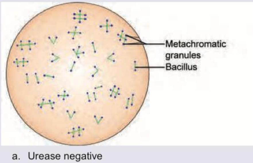
Question 3: All are true about the bacteria shown in the figure except:
- A. Pyrazinamidase test positive
- B. C. gravis has daisy head colony
- C. Heat labile toxin has affinity for myocardium
- D. Urease positive (Correct Answer)
Explanation: ***Urease positive*** - The image depicts **Corynebacterium diphtheriae**, characterized by **metachromatic granules** and its bacillary (rod) shape, often showing V or L formations. - *Corynebacterium diphtheriae* is **urease negative**, not positive, making this statement false and the correct answer to this "except" question. *Pyrazinamidase test positive* - *Corynebacterium diphtheriae* is **pyrazinamidase positive**, an important biochemical characteristic used in its identification. - This enzyme activity distinguishes it from other *Corynebacterium* species like *C. pseudodiphtheriticum* and *C. ulcerans*. *C. gravis has daisy head colony* - The **mitis** and **gravis** biotypes are distinct strains of *Corynebacterium diphtheriae*, with *C. gravis* characterized by its distinctive **"daisy head" colony morphology** on tellurite medium. - This irregular growth pattern is a known characteristic feature of *C. diphtheriae* biotype gravis. *Heat labile toxin has affinity for myocardium* - The **diphtheria toxin** produced by *C. diphtheriae* is indeed **heat-labile** and has strong affinity for various tissues, particularly the **myocardium** and nerve cells. - This tissue affinity leads to serious complications such as **myocarditis** and neurological deficits in diphtheria patients.
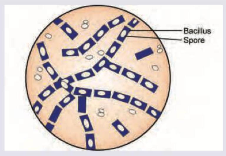
Question 4: All of the following are caused by the organism shown except:
- A. Meningitis (Correct Answer)
- B. Pneumonia
- C. Hemorrhagic diarrhea
- D. Myonecrosis
Explanation: ***Meningitis*** - The image shows **Gram-positive rods** with **spores**, characteristic of *Clostridium perfringens*. - *Clostridium perfringens* is **not a recognized cause of meningitis** in standard clinical practice. - While clostridial infections can occur in various body sites, meningitis is not among the typical clinical presentations of *C. perfringens*. - CNS infections by anaerobes typically involve other organisms like *Bacteroides* species or *Clostridium* species other than *C. perfringens*. *Hemorrhagic diarrhea* - *Clostridium perfringens* Type C causes **necrotizing enteritis (pigbel disease)** characterized by **hemorrhagic diarrhea**, particularly in areas with poor sanitation. - *C. perfringens* Type A causes **food poisoning** with watery diarrhea and abdominal cramps. - Enteritis necroticans presents with severe abdominal pain, bloody diarrhea, and can be fatal. *Pneumonia* - *Clostridium perfringens* can rarely cause **necrotizing pneumonia**, usually following aspiration or in immunocompromised patients. - This presents with rapid tissue destruction and gas formation in lung tissue. *Myonecrosis* - *Clostridium perfringens* is the **most common cause** of **gas gangrene** or **clostridial myonecrosis**. - Characterized by rapid muscle destruction due to **alpha-toxin** (lecithinase), producing gas in tissues and causing severe systemic toxicity.
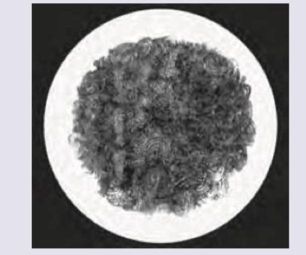
Question 5: What is the drug of choice for organism producing the following colonies?
- A. Erythromycin
- B. Ciprofloxacin (Correct Answer)
- C. Ceftriaxone
- D. No treatment with approx. 100% mortality
Explanation: ***Ciprofloxacin*** - The image displays characteristic **"Medusa head" colonies**, which are pathognomonic for *Bacillus anthracis* (anthrax). - **Ciprofloxacin** is a fluoroquinolone and is the **first-line drug of choice** for *Bacillus anthracis* infections (treatment and prophylaxis). - Other first-line options include **doxycycline**, and combination therapy is often used for systemic/inhalational anthrax. - Early antibiotic therapy significantly reduces mortality, though delayed treatment in inhalational anthrax carries high mortality risk. *Erythromycin* - Erythromycin is a **macrolide antibiotic** that is **not recommended** for *Bacillus anthracis* infections. - It has lower efficacy and is not considered effective against anthrax, especially in severe systemic forms. *Ceftriaxone* - Ceftriaxone is a **third-generation cephalosporin** that is **not recommended** for anthrax. - *Bacillus anthracis* produces **beta-lactamase enzymes** that confer resistance to many beta-lactam antibiotics. - Therefore, ceftriaxone would be ineffective as monotherapy. *No treatment with approx. 100% mortality* - This is **incorrect** as a treatment option since effective antibiotics are available. - While untreated inhalational anthrax has very high mortality (approaching 90-100%), **treatment exists and is effective**, especially when initiated early. - The drug of choice for anthrax is ciprofloxacin (or doxycycline), not "no treatment."
Question 6: What is the following pattern seen with *Clostridium perfringens* called?
- A. Nagler's reaction (Correct Answer)
- B. Reverse CAMP test
- C. Stormy clot reaction
- D. CAMP test
Explanation: ***Nagler's reaction*** - This test identifies the production of **alpha-toxin (lecithinase)** by *Clostridium perfringens*, which hydrolyzes lecithin in egg yolk agar, resulting in a **turbid halo** around the colonies. - The image shows a clear zone of inhibition (no halo) where antiserum to alpha toxin is present, confirming that the halo formation in the other section is due to the alpha toxin. *Reverse CAMP test* - The **reverse CAMP test** is used to identify *Clostridium perfringens* but involves its synergistic hemolytic effect with Group B *Streptococcus*, not the lecithinase activity directly shown here. - In this test, *Clostridium perfringens* produces an enzyme that inhibits the hemolytic activity of *Streptococcus agalactiae*, resulting in an arrow-headed zone of *no hemolysis*. *Stormy clot reaction* - The **stormy clot** is a characteristic fermentation reaction seen with *Clostridium perfringens* in **litmus milk media**, where the bacteria ferment lactose, produce gas, and coagulate the casein. - This reaction involves milk coagulation and gas production, not the lecithinase activity on egg yolk agar. *CAMP test* - The **CAMP test** detects the synergistic hemolysis between *Staphylococcus aureus* and Group B *Streptococcus (Streptococcus agalactiae)*. - It results in an **arrowhead-shaped zone of complete hemolysis** when both organisms are cultured perpendicular to each other on blood agar.
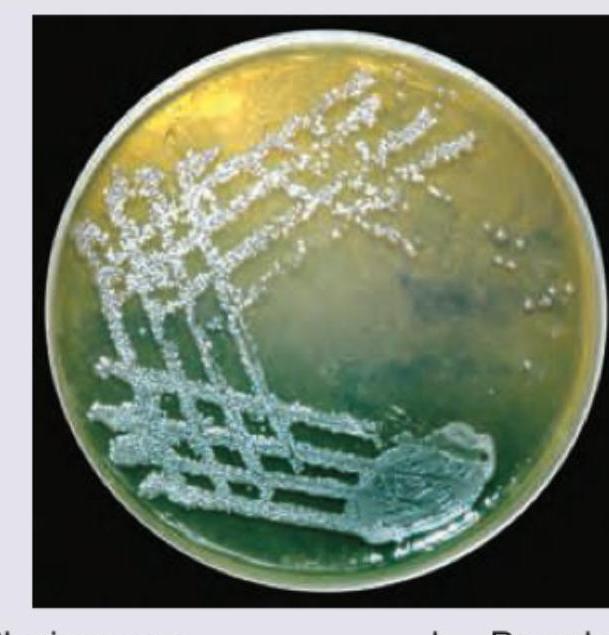
Question 7: Which of the bacteria is shown in this culture plate of nutrient agar? (Recent NEET Pattern 2016-17)
- A. Plesiomonas
- B. Pseudomonas (Correct Answer)
- C. Burkholderia
- D. Pasteurella
Explanation: ***Pseudomonas*** - The image displays colonies with a **metallic sheen** and a characteristic **greenish-blue pigment**, consistent with *Pseudomonas aeruginosa* growth on nutrient agar. - This pigmentation is due to the production of **pyocyanin** and **pyoverdin**, which are distinctive for *Pseudomonas aeruginosa*. *Plesiomonas* - *Plesiomonas shigelloides* typically does not produce the intense metallic sheen or the characteristic greenish-blue pigment seen in the image. - It is more commonly associated with aquatic environments and gastrointestinal infections, and its colonies usually appear opaque or translucent. *Burkholderia* - *Burkholderia* species can produce a variety of colony morphologies, but they generally do not exhibit the striking **metallic sheen** or the specific greenish-blue pigmentation caused by pigments like pyocyanin. - Certain *Burkholderia* species, such as *B. cepacia*, might produce yellow or green pigments, but the overall appearance in the image is more typical of *Pseudomonas*. *Pasteurella* - *Pasteurella* species typically produce smaller, gray, translucent, and non-hemolytic colonies without the distinctive **metallic sheen** or pigment seen in the culture image. - They are also facultatively anaerobic and are often associated with respiratory infections in animals and humans after animal bites.
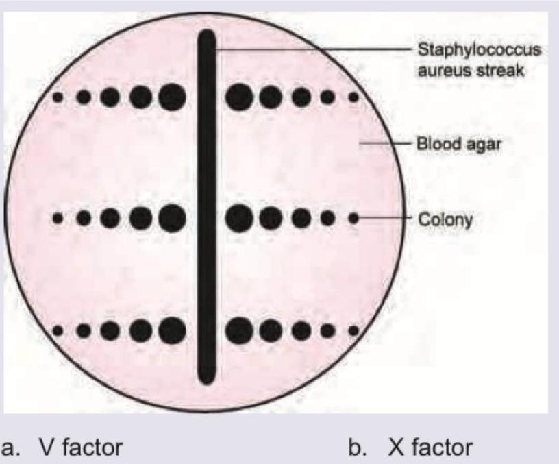
Question 8: The following plate shows *H. influenzae* colonies. Which of the following is responsible for the phenomenon shown? (Recent NEET Pattern 2016-17)
- A. V factor (Correct Answer)
- B. X factor
- C. All of the above
- D. PRP antigen
Explanation: ***V factor*** - The phenomenon depicted is **satellitism**, where *Haemophilus influenzae* grows as satellite colonies around a streak of *Staphylococcus aureus* on blood agar. - This occurs because *S. aureus* lyses red blood cells and releases **V factor (NAD - nicotinamide adenine dinucleotide)**, which is an essential growth factor that *H. influenzae* requires but cannot synthesize. - While *H. influenzae* requires both **X factor (hemin)** and **V factor (NAD)**, the satellitism phenomenon specifically demonstrates the role of **V factor** provided by *S. aureus*. - X factor is already available throughout the blood agar from lysed RBCs, so the enhanced growth specifically around *S. aureus* colonies is due to the **V factor** produced by the bacteria. *X factor* - **X factor (hemin)** is present throughout the blood agar medium from lysed red blood cells, so it does not explain the specific satellite growth pattern around *S. aureus* colonies. - Both X and V factors are required for growth, but the satellitism pattern demonstrates V factor dependency specifically. *PRP antigen* - **PRP (polyribosylribitol phosphate)** is the capsular polysaccharide of *Haemophilus influenzae* type b (Hib) and is a virulence factor. - It is not a growth factor and plays no role in the satellitism phenomenon. *All of the above* - Incorrect, as only **V factor** is responsible for the satellitism phenomenon shown in the image. - PRP antigen is not a growth factor, and X factor is already uniformly distributed in blood agar.
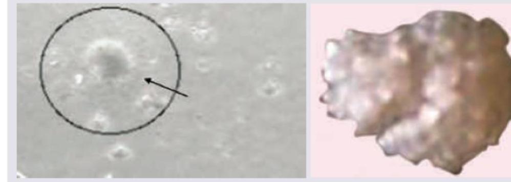
Question 9: All are true about the organism whose colonies are shown below except:
- A. Mulberry shaped colony
- B. Warm agglutinin production (Correct Answer)
- C. Lack cell wall
- D. Divide by binary fission
Explanation: ***Warm agglutinin production*** - This is the **EXCEPTION** - Mycoplasma pneumoniae produces **COLD agglutinins**, not warm agglutinins - **Cold agglutinins** are IgM antibodies that agglutinate red blood cells at temperatures below 37°C and are a characteristic feature of **Mycoplasma pneumoniae** infection - **Warm agglutinins** are associated with autoimmune hemolytic anemia, not Mycoplasma infections - This is the FALSE statement, making it the correct answer in this EXCEPT question *Mulberry shaped colony* - **TRUE statement** - Mycoplasma colonies have a characteristic **"fried egg" or mulberry appearance** with a dense central zone and a flat peripheral zone - This distinctive colony morphology is due to the organisms growing into the agar surface while spreading on top - This is a key identifying feature of Mycoplasma species on culture media *Lack cell wall* - **TRUE statement** - Mycoplasma are unique among bacteria in that they **completely lack a cell wall** - This absence of cell wall makes them resistant to **beta-lactam antibiotics** (penicillins, cephalosporins) that target cell wall synthesis - The lack of cell wall also contributes to their **pleomorphism** (variable shape) and osmotic fragility - Their cell membrane contains **sterols**, which is unusual for prokaryotes *Divide by binary fission* - **TRUE statement** - Mycoplasma reproduce by **binary fission**, the standard method of bacterial reproduction - Despite lacking a cell wall, they still undergo typical prokaryotic cell division - They are the **smallest self-replicating organisms** capable of independent growth
Question 10: A 35-year-old patient presents to the OPD 24 hours after a fight with a stranger in which he was bitten. GCS is 15/15 and following injury is noted on left forearm. He complains of extreme pain and tenderness in the injury. Swab from the injury was plated in chocolate agar and incubated in 10% carbon dioxide for 48 hours. Small colonies with pitting appearance were noted. Which of the following organism is responsible?
- A. Flavobacterium meningosepticum
- B. Capnocytophaga gingivalis
- C. Streptobacillus moniliformis
- D. Eikenella corrodens (Correct Answer)
Explanation: ***Eikenella corrodens*** - The context of a **human bite wound** and the characteristic **pitting of agar** by bacterial colonies are classic identifiers for *Eikenella corrodens*. - This organism is a common inhabitant of the **oral flora** and is frequently implicated in infections resulting from human bites. *Flavobacterium meningosepticum* - This organism is more commonly associated with **nosocomial infections**, particularly in newborns and immunocompromised patients, and severe infections like meningitis or sepsis, not typically human bite wounds. - While it can grow on chocolate agar, its colonial morphology does **not typically involve pitting** of the agar. *Capnocytophaga gingivalis* - This organism is also part of the normal oral flora and can cause infections related to human bites, especially in immunocompromised individuals. - However, while it can grow on chocolate agar, it characteristically exhibits **gliding motility** and ferments carbohydrates, but does not typically cause the striking **pitting** seen with *Eikenella corrodens*. *Streptobacillus moniliformis* - *Streptobacillus moniliformis* is associated with **rat bite fever** (Haverhill fever if contracted through contaminated food or water) and not typically human bite wounds. - It often produces **"fried egg" colonies** with a dense center and a lacy edge, which is distinct from the pitting observed here.