All SubjectsAnatomy (30)Anesthesiology (8)Biochemistry (8)Community Medicine (17)Dermatology (24)ENT (18)Forensic Medicine (18)General Medicine (2)Internal Medicine (23)Internal Medicine (8)Microbiology (39)Obstetrics and Gynecology (15)Ophthalmology (16)Orthopaedics (11)Pathology (10)Pathology (17)Pediatrics (26)Pharmacology (6)Physiology (15)Radiology (30)Surgery (5)Surgery (22)
Q11
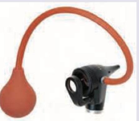
All of the following statements regarding this instrument are true except: (Recent NEET Pattern 2016-17)
Q12
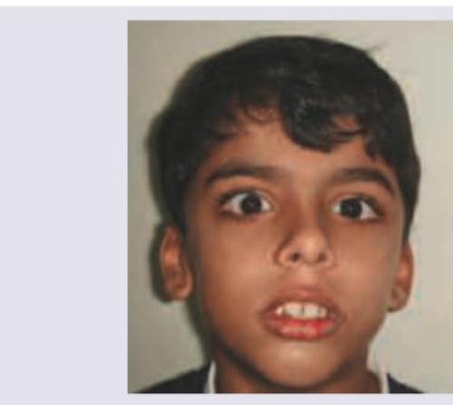
The facial features shown in the image are characteristic of:
Q13
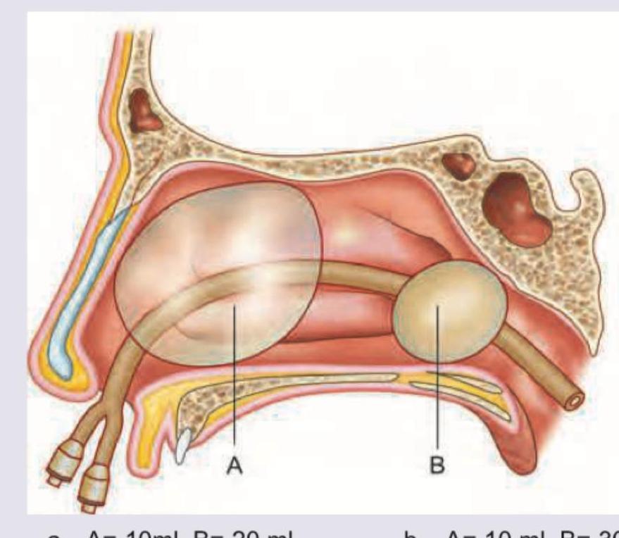
The volume of the balloons shown in epistaxis balloon is: (Recent NEET Pattern 2016-17)
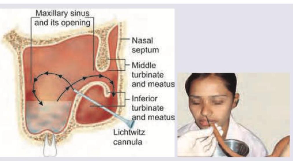
Q14
The image shows which of the following tests being performed? (Recent NEET Pattern 2016-17)
Q15
All are contraindications to the procedure shown below except:
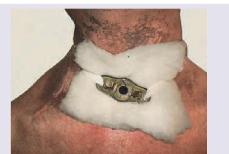
Q16
All of the following are correct about the image shown except:
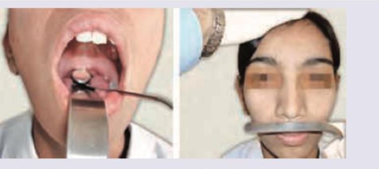
Q17
All are correct about the procedure performed in the patient except:
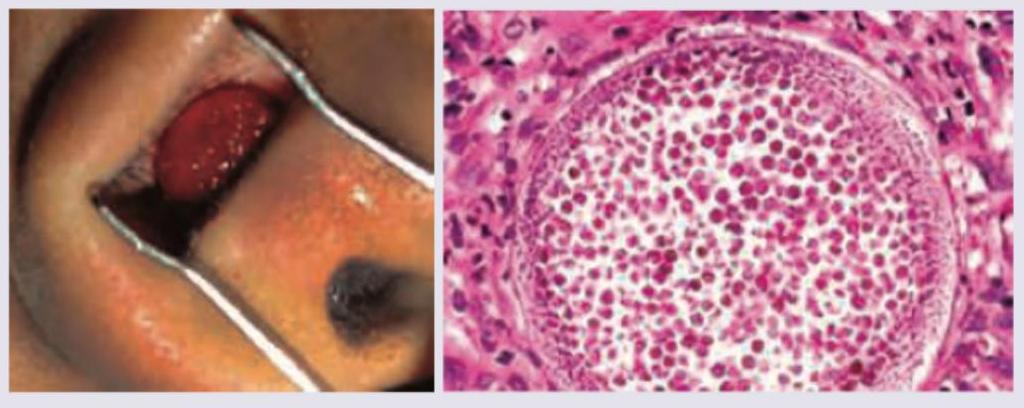
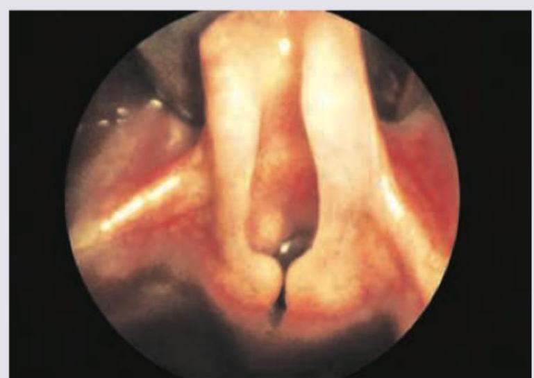
Q18
A 15-year-old female presents with nasal obstruction and occasional profuse epistaxis for last 8 weeks. Nasal speculum view and histopathology of resected lesion is given. All are correct about the diagnosis except: