NEET-PG 2017 — Dermatology
24 Previous Year Questions with Answers & Explanations
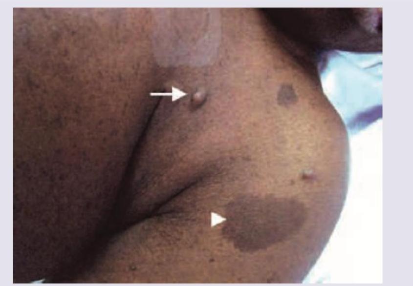
A 38-year-old man presents with the manifestation shown in the image. He has a number of family members suffering from the same condition, though the severity is different in different members. Which of the following statements is false regarding this condition?
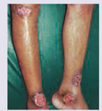
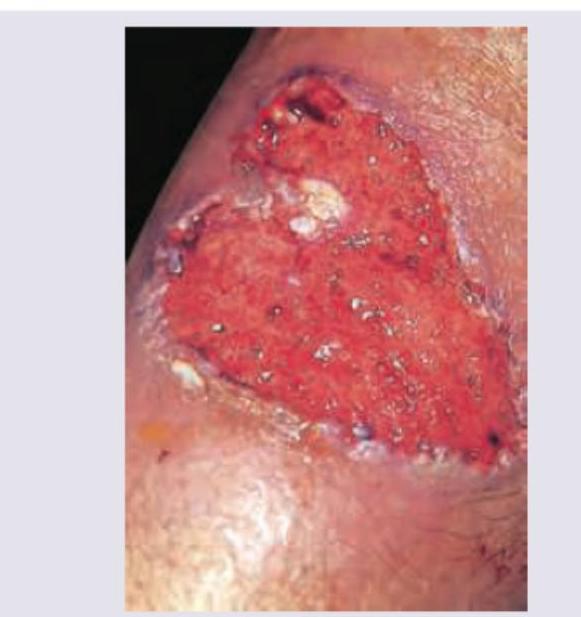
A 45-year-old Ulcerative colitis patient presents with multiple painful lesions on both legs. What is the diagnosis?
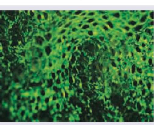
Which of the following vesico-bullous disorders will exhibit the direct immunofluorescence shown below? (Recent NEET Pattern 2016-17)
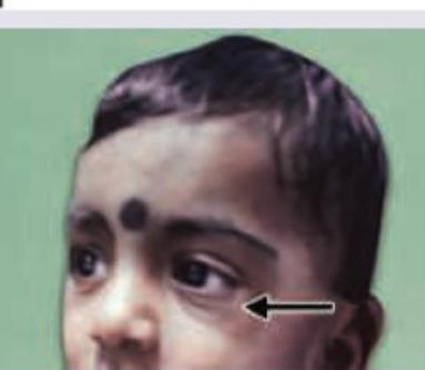
The clinical sign shown in the image is:
The following lesion appears on the leg of a patient of ulcerative colitis. All are useful in management except:
All are true about this lesion seen in a child with epilepsy except:
An 8-year-old boy who was diagnosed with hypertrophic cardiomyopathy has reddish papules all over the body developing after birth. He also complains of severe pain in hands and feet. Which of the following is correct?
All are true about the condition shown in the figure except: (Recent NEET Pattern 2016-17)
The following lesion is seen in all of the following conditions except: (Recent NEET Pattern 2016-17)
All are correct about the condition shown in the image except: (Recent NEET Pattern 2016-17)
NEET-PG 2017 - Dermatology NEET-PG Practice Questions and MCQs
Question 1: A 38-year-old man presents with the manifestation shown in the image. He has a number of family members suffering from the same condition, though the severity is different in different members. Which of the following statements is false regarding this condition?
- A. Autosomal dominant condition
- B. Chromosomal abnormality seen in chromosome 17
- C. Café-au-lait spots and neurofibromas are characteristic features
- D. Basal cell carcinoma (Correct Answer)
Explanation: ***Basal cell carcinoma*** - Basal cell carcinoma is a type of skin cancer that is **not typically a primary manifestation** of Neurofibromatosis Type 1 (NF1). - While individuals with NF1 may have an increased risk of certain cancers, basal cell carcinoma is **not one of the characteristic features** of the condition. - **This statement is FALSE**, making it the correct answer to this question. *Autosomal dominant condition* - Neurofibromatosis Type 1 (NF1) is inherited in an **autosomal dominant pattern**, meaning only one copy of the mutated gene is needed to cause the disorder. - The patient's history of **multiple family members** suffering from the same condition with varying severity is consistent with autosomal dominant inheritance. - **This statement is TRUE.** *Chromosomal abnormality seen in chromosome 17* - Neurofibromatosis Type 1 is caused by a mutation in the **NF1 gene**, located on **chromosome 17 (17q11.2)**. - This gene encodes for **neurofibromin**, a tumor suppressor protein, and its dysfunction leads to the characteristic features of NF1. - **This statement is TRUE.** *Café-au-lait spots and neurofibromas are characteristic features* - The image displays characteristic features of NF1, including **café-au-lait spots** (large, hyperpigmented macules) and **neurofibromas** (benign tumors of nerve sheath cells). - These are classic diagnostic findings in Neurofibromatosis Type 1, along with axillary/inguinal freckling and Lisch nodules. - **This statement is TRUE.**
Question 2: A 45-year-old Ulcerative colitis patient presents with multiple painful lesions on both legs. What is the diagnosis?
- A. Pyoderma gangrenosum (Correct Answer)
- B. Febrile neutropenic dermatosis
- C. Necrotizing fasciitis
- D. Granulomatosis with polyangiitis
Explanation: ***Pyoderma gangrenosum*** - This patient has **ulcerative colitis**, which is strongly associated with **pyoderma gangrenosum**, a neutrophilic dermatosis. - The image shows characteristic **painful, rapidly expanding ulcers** with violaceous, undermined borders, typical of pyoderma gangrenosum. *Febrile neutropenic dermatosis* - This condition (also known as **Sweet syndrome**) occurs in patients with **neutropenia** and **fever**, presenting with painful erythematous plaques or nodules. - While systemic illness like ulcerative colitis can predispose to skin conditions, the specific presentation and lack of mentioned neutropenia make this less likely. *Necrotizing fasciitis* - **Necrotizing fasciitis** is a rapidly progressive, life-threatening infection of the deep fascia and subcutaneous tissue, typically presenting with severe pain, erythema, swelling, and crepitus. - The lesions in the image appear to be chronic ulcers with specific borders rather than acute, rapidly spreading infection of necrotizing fasciitis. *Granulomatosis with polyangiitis* - Also known as **Granulomatosis with polyangiitis (GPA)**, formerly **Wegener's granulomatosis**, this is an autoimmune vasculitis primarily affecting the respiratory tract and kidneys, and can cause skin lesions such as palpable purpura, nodules, or ulcers. - While skin lesions can occur, the characteristic features of **pyoderma gangrenosum** and its strong association with inflammatory bowel disease make it a more probable diagnosis in this context.
Question 3: Which of the following vesico-bullous disorders will exhibit the direct immunofluorescence shown below? (Recent NEET Pattern 2016-17)
- A. Bullous pemphigoid
- B. Cicatricial pemphigoid
- C. Chronic bullous dermatosis of childhood
- D. Pemphigus vulgaris (Correct Answer)
Explanation: ***Pemphigus vulgaris*** - The direct immunofluorescence image shows a characteristic **"chicken wire"** or **intercellular** staining pattern in the epidermis. - This pattern indicates the presence of autoantibodies (typically **IgG**) targeting **desmoglein 1 and 3** in the desmosomes, leading to intraepidermal blistering, which is a hallmark of pemphigus vulgaris. *Bullous pemphigoid* - Direct immunofluorescence in bullous pemphigoid typically shows a **linear deposition of IgG and C3 along the dermal-epidermal junction** (basement membrane zone). - This pattern is distinctly different from the intercellular epidermal staining seen in the image. *Cicatricial pemphigoid* - This condition also presents with **linear deposition of immunoreactants along the basement membrane zone**, similar to bullous pemphigoid, but with a different antigen target. - The image does not show a linear basement membrane pattern, ruling out cicatricial pemphigoid. *Chronic bullous dermatosis of childhood* - Also known as **linear IgA bullous dermatosis**, it is characterized by a **linear deposition of IgA along the basement membrane zone**. - The image clearly displays an intercellular staining pattern within the epidermis, not a linear pattern at the dermal-epidermal junction.
Question 4: The clinical sign shown in the image is:
- A. Allergic salute
- B. Allergic line
- C. Dennie-Morgan fold (Correct Answer)
- D. Allergic shiners
Explanation: ***Dennie-Morgan fold (infraorbital fold)*** - The image clearly shows a prominent **infraorbital fold** or crease below the lower eyelid. - This crease is known as **Dennie-Morgan fold** (also called Dennie-Morgan lines) and is a common finding in individuals with atopic dermatitis or chronic allergic conditions. - It represents an accentuated line or fold in the skin of the lower eyelid and is considered a minor diagnostic criterion for atopic dermatitis. *Allergic salute* - An **allergic salute** refers to the characteristic gesture where a child repeatedly pushes the tip of the nose upward with the palm of the hand to relieve nasal itching and obstruction. - This action often leads to a transverse crease across the bridge of the nose, but it is not depicted in the image. *Allergic line* - The term **allergic line** (or allergic crease) is synonymous with the transverse nasal crease resulting from the allergic salute. - While it's a sign associated with chronic allergic rhinitis, it does not describe the infraorbital fold seen in the image. *Allergic shiners* - **Allergic shiners** are dark, discolored areas under the eyes, resembling bruises, caused by venous congestion secondary to chronic allergic rhinitis or nasal obstruction. - While allergic shiners may coexist with Dennie-Morgan folds, the prominent feature indicated in the image is the infraorbital fold itself, not periorbital darkening.
Question 5: The following lesion appears on the leg of a patient of ulcerative colitis. All are useful in management except:
- A. Steroids
- B. Sulfapyridine (Correct Answer)
- C. Procto-colectomy
- D. Infliximab
Explanation: ***Sulfapyridine*** - The image shows **pyoderma gangrenosum**, a painful ulcerative skin condition often associated with inflammatory bowel disease like ulcerative colitis. Among the given options, **sulfapyridine** has the **least established role** in pyoderma gangrenosum management. - **Sulfapyridine** is an inactive component of **sulfasalazine** and primarily acts as an **antibacterial agent**. While sulfasalazine has been reported in some PG cases, sulfapyridine alone is not a recognized treatment for the inflammatory, non-infectious nature of pyoderma gangrenosum. - Unlike the other options which have well-established roles, sulfapyridine lacks strong evidence for efficacy in PG. *Steroids* - **Corticosteroids** (oral or topical) are the **first-line treatment** for pyoderma gangrenosum due to their potent anti-inflammatory and immunosuppressive effects. - They help to reduce the inflammation and promote healing of the painful ulcers. *Procto-colectomy* - In cases of severe, refractory pyoderma gangrenosum associated with ulcerative colitis, **colectomy** can be a **definitive treatment** as it removes the underlying inflammatory trigger. - This surgical intervention is considered when medical therapies are unsuccessful or when the colonic disease itself necessitates surgery. *Infliximab* - **Infliximab**, a **TNF-alpha inhibitor**, is a biologic agent effective in treating both ulcerative colitis and pyoderma gangrenosum. - It is used in cases that are refractory to steroids or when patients cannot tolerate steroid therapy.
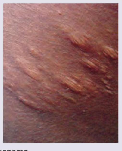
Question 6: All are true about this lesion seen in a child with epilepsy except:
- A. Collagenoma
- B. Minor criteria for diagnosis (Correct Answer)
- C. Peau d'orange appearance
- D. Predominantly seen over trunk
Explanation: ***Minor criteria for diagnosis*** - The presented lesion is a **Shagreen patch**, which is considered a **major diagnostic criterion** for **Tuberous Sclerosis Complex (TSC)**, not a minor one. - A definitive diagnosis of TSC requires two major criteria or one major and two minor criteria. *Collagenoma* - A Shagreen patch is a type of dermal **collagenoma**, characterized by an overgrowth of connective tissue, primarily collagen. - These lesions often feel like **roughened or leathery plaques** on the skin. *Peau d'orange appearance* - The Shagreen patch is often described as having a **'peau d'orange'** or orange peel-like texture due to its irregular surface. - This characteristic texture helps in its clinical identification. *Predominantly seen over trunk* - Shagreen patches are typically located on the **trunk**, especially in the lumbosacral region, as seen in the image. - They are one of the distinctive cutaneous manifestations of TSC.
Question 7: An 8-year-old boy who was diagnosed with hypertrophic cardiomyopathy has reddish papules all over the body developing after birth. He also complains of severe pain in hands and feet. Which of the following is correct?
- A. Angiokeratoma, Fabry's disease (Correct Answer)
- B. Actinic keratosis, Xeroderma pigmentosa
- C. Pseudoxanthomas, pseudoxanthoma elasticum
- D. Angiofibroma, tuberous sclerosis
Explanation: ***Angiokeratoma, Fabry's disease*** - The constellation of **hypertrophic cardiomyopathy**, **reddish papules (angiokeratomas)**, and **severe pain in hands and feet (acroparesthesias)** are classic symptoms of **Fabry's disease**. - Fabry's disease is an **X-linked lysosomal storage disorder** caused by a deficiency of **alpha-galactosidase A**, leading to the accumulation of globotriaosylceramide (Gb3). *Actinic keratosis, Xeroderma pigmentosa* - **Actinic keratosis** are precancerous skin lesions caused by sun exposure, typically appearing in older adults, not as widespread papules from birth in a child. - **Xeroderma pigmentosum** is characterized by extreme sensitivity to UV light, leading to a high incidence of skin cancers, freckle-like pigmentation, and ocular abnormalities, but not typically hypertrophic cardiomyopathy or these specific papules. *Pseudoxanthomas, pseudoxanthoma elasticum* - **Pseudoxanthoma elasticum** causes small, yellowish papules, mainly on flexible skin areas, along with angioid streaks in the retina and cardiovascular issues due to calcification of elastic fibers. - While it can involve the heart, it doesn't typically present with the widespread reddish papules (angiokeratomas) or the severe acroparesthesias described. *Angiofibroma, tuberous sclerosis* - **Angiofibromas** are facial papules associated with **tuberous sclerosis**, which is also characterized by brain tubers, renal angiomyolipomas, and hypomelanotic macules. - While tuberous sclerosis can cause cardiac rhabdomyomas (tumors), it is not typically associated with widespread angiokeratomas or acroparesthesias as seen in this case.
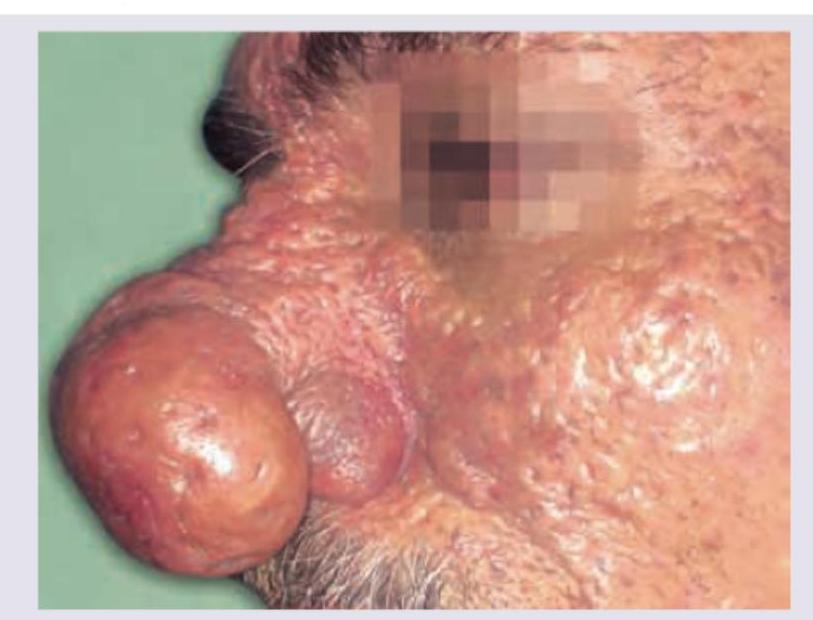
Question 8: All are true about the condition shown in the figure except: (Recent NEET Pattern 2016-17)
- A. Interconnecting sinus tracts (Correct Answer)
- B. Patulous pilo-sebaceous orifices
- C. Foul smelling cheesy material
- D. Can lead to difficulty in breathing
Explanation: ***Interconnecting sinus tracts*** - This is a characteristic feature of **Acne Conglobata**, NOT rhinophyma. Acne conglobata is a severe form of nodulocystic acne characterized by multiple interconnected comedones, abscesses, cysts, and draining sinus tracts, typically affecting the trunk, face, and neck. - **Rhinophyma** is a severe manifestation of rosacea involving progressive hypertrophy of sebaceous glands and connective tissue of the nose, producing a bulbous, enlarged appearance. It does **not** feature interconnecting sinus tracts. - This is the FALSE statement about rhinophyma, making it the correct answer for this "except" question. *Patulous pilo-sebaceous orifices* - This is a **hallmark feature of rhinophyma**. The sebaceous gland hyperplasia leads to markedly **dilated follicular openings** (patulous orifices) on the nasal surface. - These prominent, enlarged pores are a key diagnostic sign and contribute to the characteristic cobblestone appearance of the affected nose. *Foul smelling cheesy material* - The hypertrophied sebaceous glands in **rhinophyma** produce excessive sebum which accumulates in the dilated follicular openings. - This material consists of **keratin plugs, sebum, and bacterial debris**, often presenting as a foul-smelling, cheesy substance that can be expressed from the enlarged pores. *Can lead to difficulty in breathing* - **TRUE for severe rhinophyma**. Progressive nasal tissue hypertrophy can cause **external nasal valve obstruction** and narrowing of the nasal airways, leading to breathing difficulties. - Severe cases may require **surgical intervention** (e.g., laser therapy, surgical excision) not only for cosmetic reasons but also to relieve nasal obstruction and improve airflow. - This is a recognized complication documented in dermatology and otolaryngology literature.
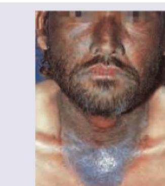
Question 9: The following lesion is seen in all of the following conditions except: (Recent NEET Pattern 2016-17)
- A. Carcinoid syndrome
- B. Hartnup disease
- C. Alcoholism
- D. Porphyria (Correct Answer)
Explanation: ***Correct: Porphyria*** - **Porphyria** causes **photosensitive skin lesions** but does NOT cause pellagra or the **Casal's necklace** pattern shown in the image. - Porphyria results from defects in **heme synthesis**, leading to vesicles, bullae, and hyperpigmentation, particularly on sun-exposed areas. - The image depicts **Pellagra**, characterized by **photosensitive dermatosis** in a **Casal's necklace** distribution, caused by **niacin (vitamin B3) deficiency**. *Incorrect: Carcinoid syndrome* - **Carcinoid syndrome** can cause **pellagra** due to **tryptophan diversion** for excessive serotonin synthesis. - This leads to **niacin deficiency** and can present with the characteristic skin lesions shown in the image. - Classic features include flushing, diarrhea, and cardiac involvement. *Incorrect: Hartnup disease* - **Hartnup disease** is an **autosomal recessive disorder** with impaired absorption of neutral amino acids, including **tryptophan**. - Reduced tryptophan absorption leads to **secondary niacin deficiency**, causing pellagra-like symptoms. - Patients present with photosensitive dermatitis, cerebellar ataxia, and psychiatric manifestations. *Incorrect: Alcoholism* - **Chronic alcoholism** is a common cause of **pellagra** due to poor dietary intake, malabsorption, and impaired nutrient metabolism. - Alcoholics are at high risk for **niacin deficiency**, presenting with the **3 Ds**: dermatitis, diarrhea, and dementia. - The photosensitive rash in a Casal's necklace distribution is characteristic.
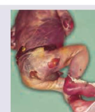
Question 10: All are correct about the condition shown in the image except: (Recent NEET Pattern 2016-17)
- A. Epidermolysin mediated
- B. Staphylococcal scalded skin syndrome
- C. Caused by gram positive cocci in clusters
- D. Necrotising Fasciitis (Correct Answer)
Explanation: ***Necrotising Fasciitis*** - The image shows widespread superficial blistering and peeling of the skin, resembling a burn, which is characteristic of **Staphylococcal Scalded Skin Syndrome (SSSS)**, not necrotizing fasciitis. - **Necrotizing fasciitis** is a deeper, rapidly spreading infection of the subcutaneous tissue and fascia, characterized by severe pain, dusky skin, bullae, and crepitus, which is not depicted here. *Epidermolysin mediated* - **Staphylococcal Scalded Skin Syndrome (SSSS)** is indeed caused by **exotoxins (epidermolysins A and B)** produced by *Staphylococcus aureus* strains, which target desmoglein 1, leading to widespread epidermal detachment. - These epidermolysins act as **superantigens**, causing a systemic toxic effect without direct bacterial invasion of the skin at affected sites. *Staphyloccocal scalded skin syndrome* - The image is pathognomonic for **Staphylococcal Scalded Skin Syndrome (SSSS)**, characterized by generalized erythema, flaccid blisters, and epidermal exfoliation, particularly in infants and young children. - The peeling skin suggests a **positive Nikolsky sign**, where slight rubbing of the skin causes the epidermis to separate from the dermis, a hallmark of SSSS. *Caused by gram positive cocci in clusters* - **Staphylococcal Scalded Skin Syndrome (SSSS)** is caused by specific strains of **Gram-positive cocci in clusters**, namely *Staphylococcus aureus*, which produce exfoliative toxins. - These bacteria are commonly found on the skin and mucous membranes and are responsible for a variety of skin and soft tissue infections.