NEET-PG 2017 — Biochemistry
6 Previous Year Questions with Answers & Explanations
A patient reports a change in colour of urine on air exposure. All are true about the condition shown below except:
All are correct about the amino acid marked as X, EXCEPT:
Name the plot dealing with kinetics of enzyme inhibition?
Which enzyme marked as $X$ is missing in synthesis of vitamin $D_{3}$ ?
The following reaction occurs in which part of kidney?
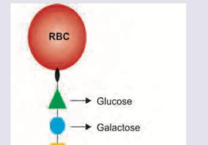
Name the antigen marked as X determining blood group A.
NEET-PG 2017 - Biochemistry NEET-PG Practice Questions and MCQs
Question 1: A patient reports a change in colour of urine on air exposure. All are true about the condition shown below except:
- A. Blackening of urine is accelerated on exposure to sunlight
- B. Alkaptone bodies are deposited in intervertebral disc
- C. Urine Benedict's test is negative (Correct Answer)
- D. The condition is caused by deficiency of homogentisate 1,2-dioxygenase
Explanation: ***Urine Benedict's test is negative*** - This is FALSE - Benedict's test is actually **POSITIVE** in alkaptonuria because **homogentisic acid** is a reducing agent. - Homogentisic acid readily **reduces Benedict's reagent**, giving a positive test result in alkaptonuria patients. *Blackening of urine is accelerated on exposure to sunlight* - This is TRUE - **UV light** and sunlight accelerate the **oxidation of homogentisic acid** in urine. - The characteristic **dark discoloration** occurs more rapidly when exposed to light and air. *Alkaptone bodies are deposited in intervertebral disc* - This is TRUE - **Homogentisic acid (alkaptone bodies)** polymerizes to form **ochronotic pigment** deposits. - These deposits accumulate in **cartilage** including intervertebral discs, causing degenerative changes and spondylosis. *The condition is caused by deficiency of homogentisate 1,2-dioxygenase* - This is TRUE - Alkaptonuria is caused by deficiency of **homogentisate 1,2-dioxygenase** enzyme in the **tyrosine metabolic pathway**. - This enzyme deficiency leads to accumulation of **homogentisic acid** in blood and urine, causing the characteristic symptoms.
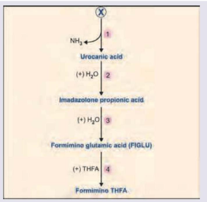
Question 2: All are correct about the amino acid marked as X, EXCEPT:
- A. Maximum buffering action
- B. Ketogenic (Correct Answer)
- C. Contributes to one carbon pool
- D. Excess amount leads to mental retardation
Explanation: ***Correct Answer: Ketogenic*** ✓ - Histidine is **purely glucogenic**, NOT ketogenic - Its carbon skeleton is converted to α-ketoglutarate (a TCA cycle intermediate), which can be used for gluconeogenesis - It does NOT produce acetyl-CoA or acetoacetate (ketone body precursors) - **This statement is FALSE, making it the correct answer for this EXCEPT question** *Incorrect: Maximum buffering action* - This statement is TRUE (so not the answer) - Histidine contains an **imidazole ring** with pKa ~6.0, close to physiological pH - Provides crucial **buffering capacity** in blood and tissues - Most effective amino acid buffer at physiological pH *Incorrect: Contributes to one carbon pool* - This statement is TRUE (so not the answer) - Histidine breakdown produces **Formimino glutamic acid (FIGLU)** - FIGLU donates its formimino group to **Tetrahydrofolate (THF)** - Contributes to the **one-carbon pool** essential for biosynthetic pathways *Incorrect: Excess amount leads to mental retardation* - This statement is TRUE (so not the answer) - **Histidinemia** results from histidase enzyme deficiency - Elevated histidine levels can be associated with **developmental delays and speech defects** in some cases - Though many affected individuals remain asymptomatic
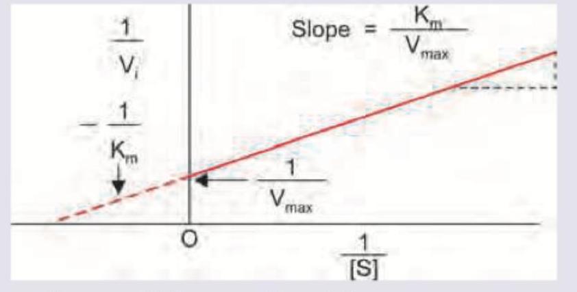
Question 3: Name the plot dealing with kinetics of enzyme inhibition?
- A. Donnan-Gibbs equation plot
- B. Lineweaver-Burk plot
- C. Dixon plot (Correct Answer)
- D. Hanes-Woolf plot
Explanation: ***Dixon plot*** - The **Dixon plot** is specifically used to determine the type of enzyme inhibition and to calculate the **inhibition constant (Ki)**. - It plots the reciprocal reaction velocity (1/V) against the **inhibitor concentration ([I])** at different substrate concentrations. *Donnan-Gibbs equation plot* - The **Donnan-Gibbs equation** describes the distribution of charged particles across a semi-permeable membrane at equilibrium. - It is irrelevant to enzyme kinetics or inhibition. *Lineweaver-Burk plot* - While the Lineweaver-Burk plot is used in enzyme kinetics to determine **Km** and **Vmax** and analyze inhibition, it plots **1/V against 1/[S]**. - The image provided, showing 1/V against [I], is characteristic of a Dixon plot, not a Lineweaver-Burk plot which plots 1/V against 1/[S]. *Hanes-Woolf plot* - The **Hanes-Woolf plot** is another linearization of the Michaelis-Menten equation, plotting **[S]/V against [S]**. - Like the Lineweaver-Burk plot, it is used for analyzing general enzyme kinetics but not specifically for determining Ki from varying inhibitor concentrations in the manner shown.
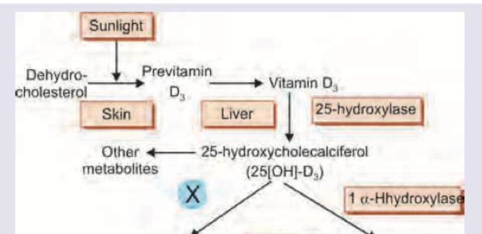
Question 4: Which enzyme marked as $X$ is missing in synthesis of vitamin $D_{3}$ ?
- A. 3-beta-hydroxylase
- B. 17-alpha-hydroxylase (Correct Answer)
- C. 21-hydroxylase
- D. 24-hydroxylase
Explanation: ***17-alpha-hydroxylase*** - The diagram illustrates the synthesis of **vitamin D3**, which involves hydroxylation steps at positions C-25 in the liver and C-1 in the kidneys. These are catalyzed by **25-hydroxylase** and **1-alpha-hydroxylase**, respectively. - **17-alpha-hydroxylase** is involved in the synthesis of steroid hormones (like cortisol, androgens, and estrogens) from cholesterol, not in the synthesis or metabolism of vitamin D3; therefore, its absence would not be relevant to vitamin D synthesis. *3-beta-hydroxylase* - **3-beta-hydroxysteroid dehydrogenase** is crucial for the synthesis of all steroid hormones, converting delta-5-3-hydroxysteroids to delta-4-3-ketosteroids. - Its presence is essential for **steroidogenesis**, but it does not play a direct role in the specific hydroxylation steps for vitamin D activation. *21-hydroxylase* - **21-hydroxylase** is an enzyme involved in the synthesis of steroid hormones such as cortisol and aldosterone from progesterone and 17-hydroxyprogesterone. - Deficiency of this enzyme leads to **congenital adrenal hyperplasia**, a condition unrelated to vitamin D metabolism. *24-hydroxylase* - **24-hydroxylase** is responsible for the inactivation of vitamin D metabolites by adding a hydroxyl group at position C-24, leading to degradation. - This enzyme is part of the **catabolic pathway** to regulate vitamin D levels, rather than being a missing enzyme in the primary synthesis of active vitamin D.
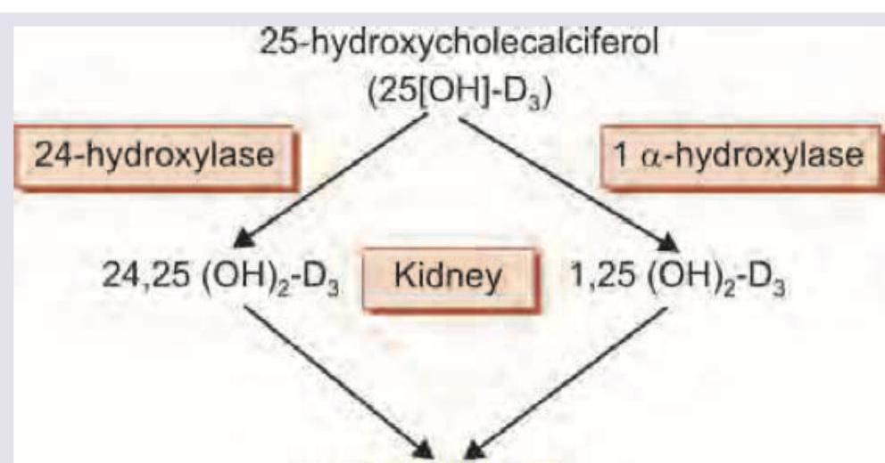
Question 5: The following reaction occurs in which part of kidney?
- A. Proximal convoluted tubule (Correct Answer)
- B. Distal convoluted tubule
- C. Loop of Henle
- D. Collecting duct
Explanation: **Proximal convoluted tubule** - The image shows the conversion of 25-hydroxycholecalciferol to **1,25 (OH)₂-D₃**, also known as calcitriol, via the enzyme **1α-hydroxylase**. - This critical hydroxylation reaction, occurring primarily in the **proximal convoluted tubule** cells of the kidney, produces the biologically active form of vitamin D. *Distal convoluted tubule* - The distal convoluted tubule is primarily involved in **fine-tuning** water and electrolyte reabsorption, influenced by hormones like aldosterone and antidiuretic hormone. - It does not contain the necessary enzymes, specifically **1α-hydroxylase**, for the final activation step of vitamin D. *Loop of Henle* - The loop of Henle's main function is to create a **medullary osmotic gradient** through countercurrent multiplication, crucial for concentrating urine. - It plays no significant role in the **hydroxylation of vitamin D** precursors. *Collecting duct* - The collecting duct is responsible for final adjustments to urine volume and concentration, largely under the influence of **antidiuretic hormone**. - It lacks the **enzymatic machinery** (1α-hydroxylase) required for the activation of vitamin D.
Question 6: Name the antigen marked as X determining blood group A.
- A. N-Acetyl-Glucosamine
- B. Dermatan sulphate
- C. Keratan sulfate
- D. N-Acetyl-Galactosamine (Correct Answer)
Explanation: ***N-Acetyl-Galactosamine*** - Blood group A antigens are formed by the addition of **N-acetylgalactosamine** to the H antigen precursor molecule on the surface of red blood cells. - This sugar modification is catalyzed by the **A transferase enzyme**, which is specific for N-acetylgalactosamine. *N-Acetyl-Glucosamine* - While N-acetylglucosamine is a component of many glycans, it is not the terminal sugar that defines the **blood group A antigen**. - **N-acetylglucosamine** is a key building block for the H antigen and other blood group precursors, but not the specific modifying sugar for A. *Dermatan sulphate* - **Dermatan sulfate** is a **glycosaminoglycan** primarily found in connective tissues, skin, and blood vessels. - It plays a role in wound healing and coagulation, but is not involved in **ABO blood group determination**. *Keratan sulfate* - **Keratan sulfate** is another **glycosaminoglycan** found in cartilage, cornea, and bone. - It contributes to tissue hydration and structural integrity, but it is not part of the **ABO blood group antigens**.