Which nerve marked as X innervates the Aortic Arch?
Which of the following is correct about the type of neuron shown below?
In the sarcomere diagram shown below, what do the marked areas X and Y represent?
Which of the following area of visual cortex is related to color vision?
Name the pathways marked as $X$ and $Y$.
NEET-PG 2017 - Anatomy NEET-PG Practice Questions and MCQs
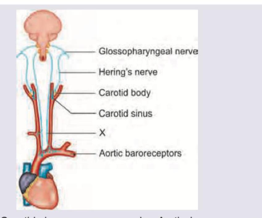
Question 31: Which nerve marked as X innervates the Aortic Arch?
- A. Carotid sinus nerve
- B. Inferior cervical cardiac nerve
- C. Superior cervical cardiac nerve
- D. Aortic depressor nerve (Correct Answer)
Explanation: ***Aortic depressor nerve*** - The nerve marked as X in the diagram directly innervates the **aortic baroreceptors** located in the aortic arch. - This nerve is also known as the **aortic depressor nerve**, a branch of the vagus nerve (CN X), which transmits sensory information about blood pressure from the aortic arch to the central nervous system. *Carotid sinus nerve* - The carotid sinus nerve (also known as Hering's nerve) innervates the **carotid sinus and carotid body**, which are located at the bifurcation of the common carotid artery. - This nerve transmits sensory information from the carotid baroreceptors and chemoreceptors, distinct from the aortic arch. *Inferior cervical cardiac nerve* - The inferior cervical cardiac nerve is a **sympathetic nerve** that originates from the inferior cervical ganglion and innervates the heart. - It does not primarily innervate the aortic arch baroreceptors; its function is related to cardiac rate and contractility. *Superior cervical cardiac nerve* - Similar to the inferior cervical cardiac nerve, the superior cervical cardiac nerve is a **sympathetic nerve** originating from the superior cervical ganglion. - It primarily contributes to the cardiac plexus and innervates the heart, not specifically the aortic arch baroreceptors.
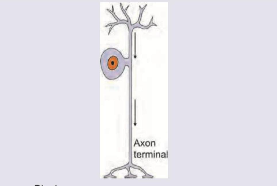
Question 32: Which of the following is correct about the type of neuron shown below?
- A. Bipolar neuron
- B. Unipolar cell
- C. Pseudo-unipolar cell (Correct Answer)
- D. All of the above
Explanation: ***Pseudo-unipolar cell*** - The image shows a neuron with a **single process** emerging from the cell body that then **divides into two branches** (one leading to dendrites and the other to the axon terminal), which is characteristic of a pseudo-unipolar neuron. - These neurons are typically found in **sensory ganglia**, such as the dorsal root ganglia, where they transmit sensory information. *Bipolar neuron* - A bipolar neuron has **two distinct processes** extending from the cell body: one axon and one dendrite. - Examples include neurons found in the **retina** and **olfactory epithelium**. *Unipolar cell* - A unipolar neuron has a **single process** extending from the cell body, which serves as both the dendrite and the axon. - These are typically found in **invertebrates**, though the term can sometimes be confusingly used for pseudo-unipolar neurons in some contexts. *All of the above* - This is incorrect because the neuron depicted specifically matches the morphology of a pseudo-unipolar cell, not all the listed types. - Each neuron type has distinct morphological features (number of poles/processes from the cell body).
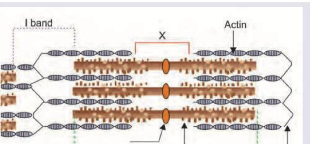
Question 33: In the sarcomere diagram shown below, what do the marked areas X and Y represent?
- A. X=H zone, Y=A band (Correct Answer)
- B. X=A band, Y=H band
- C. X=Z line, Y=M line
- D. X=M line, Y=Z line
Explanation: ***X=H zone, Y=A band*** - **X** points to the central region of the A band, visible only in a relaxed sarcomere, which is called the **H zone**, containing only thick myosin filaments. - **Y** encompasses the entire length of the thick filaments, including the regions where they overlap with thin filaments, defining the **A band**. *X=A band, Y=H band* - This is incorrect because X specifically indicates the central, lighter region within the A band, which is the H zone. - Y points to the entire segment occupied by the thick filaments, which is the A band. *X=Z line, Y=M line* - The **Z line** marks the boundaries of a sarcomere, anchoring the thin (actin) filaments, and is not indicated by X. - The **M line** is the central line within the H zone that anchors the thick (myosin) filaments, and is not indicated by Y. *X=M line, Y=Z line* - As explained, X indicates the **H zone**, which is a broader region than the M line. - Y indicates the **A band**, and not the Z line; the Z line is located at the ends of the sarcomere.
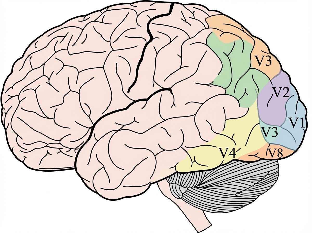
Question 34: Which of the following area of visual cortex is related to color vision?
- A. V1
- B. V8 (Correct Answer)
- C. V2
- D. V3
Explanation: ***V8*** - **V8** is a specific area within the human **visual cortex** that has been implicated in **color perception**. - Damage to this area can lead to **cerebral achromatopsia**, a condition where individuals lose the ability to perceive colors. *V1* - **V1**, also known as the **primary visual cortex**, processes basic visual information such as orientation, spatial frequency, and color. - While it processes color information, it is not considered the primary or most specialized area for **color vision** compared to V8. *V2* - **V2** receives input from V1 and is involved in processing more complex visual features, including **form, depth, and color**. - It plays a role in color processing but is less specialized for this function than V8. *V3* - **V3** is primarily involved in processing **dynamic form** and motion, with some contribution to complex visual features. - It is not extensively associated with **color perception** as its main function.
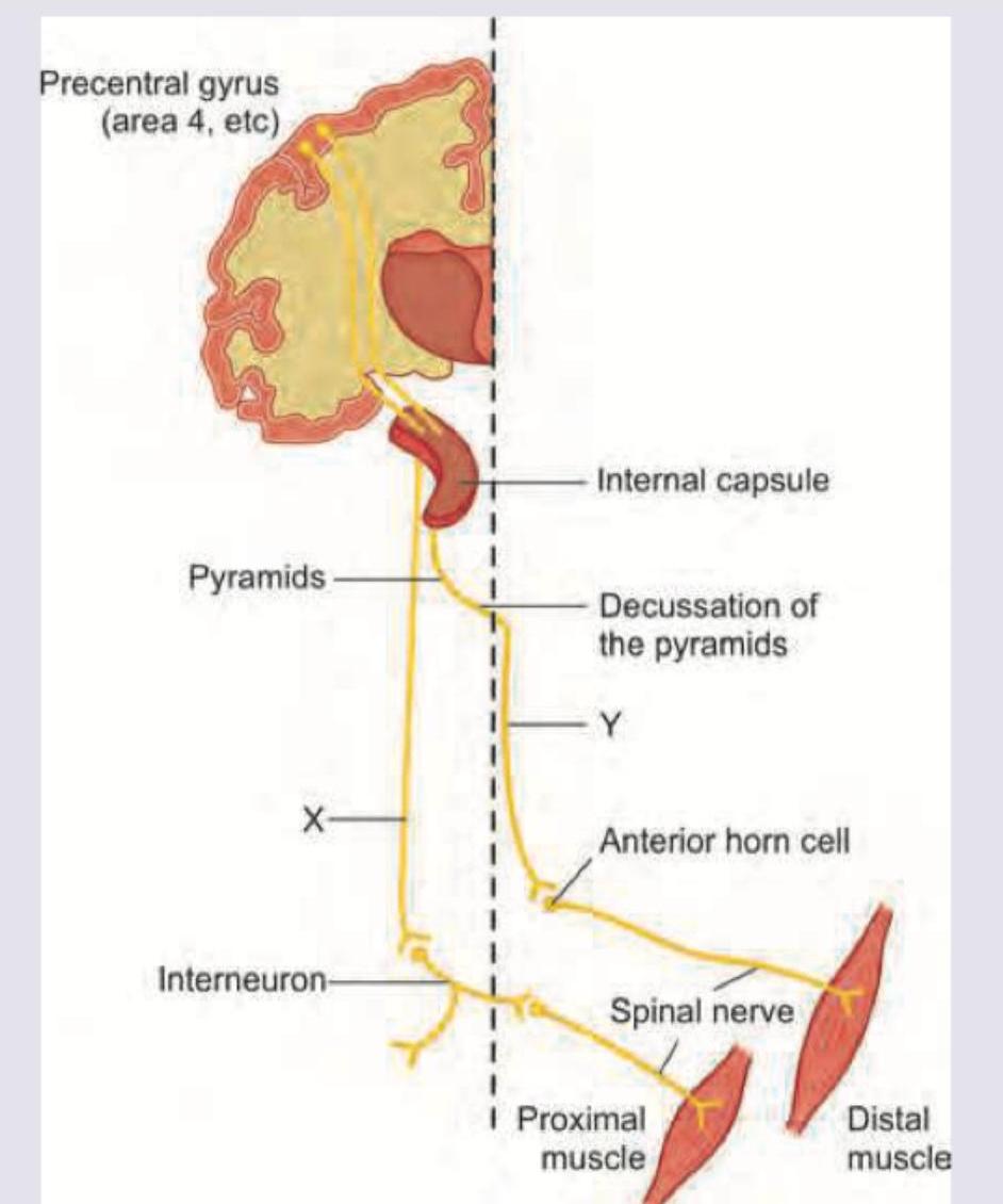
Question 35: Name the pathways marked as $X$ and $Y$.
- A. $X=$ Ventral corticospinal pathway and $Y=$ Lateral spinothalamic Pathway
- B. $X=$ Ventral corticospinal pathway and $Y=$ Lateral corticospinal pathway (Correct Answer)
- C. $X=$ Ventral corticospinal pathway and $Y=$ Lateral spinocerebellar pathway
- D. $X=$ Lateral corticospinal pathway and $Y=$ Ventral corticospinal pathway
Explanation: ***X = Ventral corticospinal pathway and Y = Lateral corticospinal pathway*** - The diagram illustrates the **corticospinal tracts**, which control voluntary movement. Pathway Y shows fibers descending from the cortex, **decussating** (crossing over) at the pyramids, and then continuing down the contralateral side to innervate distal muscles, characteristic of the **lateral corticospinal tract**. - Pathway X shows fibers that descend **ipsilaterally** (on the same side) from the cortex, then decussate at the spinal cord level to innervate proximal muscles, which is typical for the **ventral (anterior) corticospinal tract**. *X = Ventral corticospinal pathway and Y = Lateral spinothalamic Pathway* - The **lateral spinothalamic pathway** is an ascending sensory pathway for pain and temperature, originating in the spinal cord and ascending to the thalamus, rather than a descending motor pathway as shown by Y. - The pathways shown (X and Y) are clearly originating from the motor cortex (precentral gyrus) and descending to muscles, indicating they are **motor pathways**, not sensory. *X = Ventral corticospinal pathway and Y = Lateral spinocerebellar pathway* - The **lateral spinocerebellar pathway** is predominantly an ascending pathway carrying unconscious proprioceptive information to the cerebellum, not a descending motor pathway synapsing on lower motor neurons for voluntary muscle control. - Pathway Y is shown forming synapses with **anterior horn cells** controlling skeletal muscles, indicating it is a part of the motor system originating from the precentral gyrus. *X = Lateral corticospinal pathway and Y = Ventral corticospinal pathway* - This option incorrectly identifies pathway X as lateral and Y as ventral. The diagram clearly shows that pathway Y crosses over at the level of the pyramids (medulla) to descend on the contralateral side, which is the defining characteristic of the **lateral corticospinal pathway**. - Pathway X descends Ipsilaterally and crosses at segmental levels in the spinal cord, which is characteristic of the **ventral (anterior) corticospinal pathway**.