INI-CET 2023 — Physiology
11 Previous Year Questions with Answers & Explanations
JAK-STAT pathway is seen in which of the following?
Absence or mutation of SRY gene results in ?
What is the order of bands in a sarcomere from the Z-disc toward the center?
The bitter taste of toxic substances prevents us from their consumption, which of the following elicits the bitterness?
Windkessel effect is not shown by which of the following vessel?
Cystic fibrosis leads to defect in which of the following channels?
Find the correct Auditory pathway sequence.
Arrange the following parts of sarcomere from periphery to center. 1. Z line 2. M line 3. A band 4. H zone
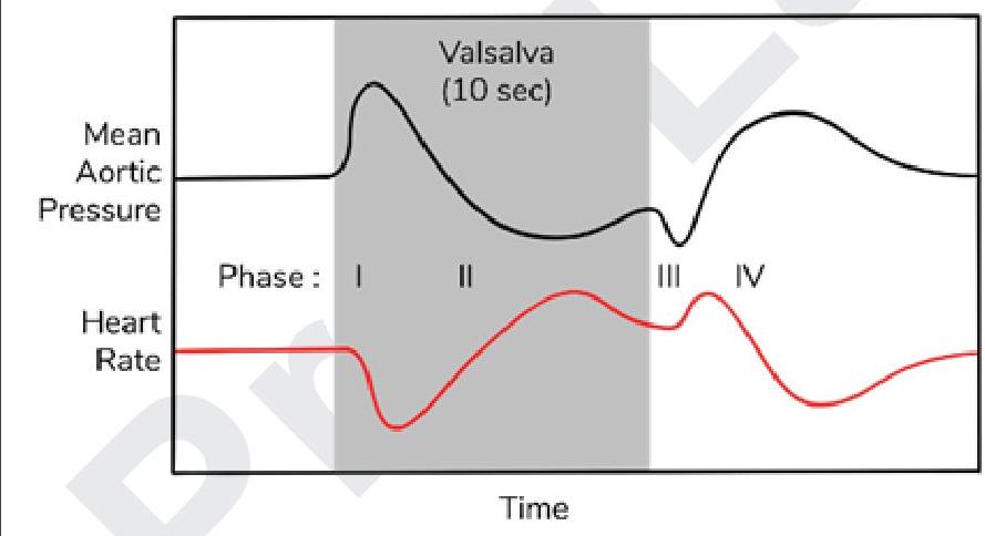
Blood pressure changes in radial artery were measured. Which of the following is the reason for initial rise in BP while performing Valsalva maneuver?
Assertion: RMP depends on proteins and phosphate ions. Reason: Diffusion potential can be calculated using nernst equation. Choose the best statement regarding the assertion and reason.
INI-CET 2023 - Physiology INI-CET Practice Questions and MCQs
Question 1: JAK-STAT pathway is seen in which of the following?
- A. Calcitonin
- B. Aldosterone
- C. Vasopressin
- D. Leptin (Correct Answer)
Explanation: ***Leptin*** - **Leptin** binding to its receptor activates the **JAK-STAT pathway**, regulating appetite and metabolism. - This pathway involves the phosphorylation of **STAT proteins**, which then translocate to the nucleus to induce gene expression. *Calcitonin* - **Calcitonin** activates **G protein-coupled receptors**, leading to an increase in intracellular cyclic AMP (cAMP). - Its primary role is in **calcium homeostasis**, lowering blood calcium levels. *Aldosterone* - **Aldosterone** is a steroid hormone that binds to **intracellular mineralocorticoid receptors**. - This complex then acts as a **transcription factor**, affecting gene expression in the kidneys to regulate sodium and potassium balance. *Vasopressin* - **Vasopressin** (ADH) binds to **G protein-coupled receptors** (V1 and V2 receptors). - V2 receptor activation in the kidney leads to increased **cAMP** and insertion of aquaporins, regulating water reabsorption.
Question 2: Absence or mutation of SRY gene results in ?
- A. Hydrocele testis
- B. Undescended testis
- C. None of the options
- D. Gonadal dysgenesis (Correct Answer)
Explanation: ***Gonadal dysgenesis*** - The **SRY gene** (Sex-determining region Y gene) is critical for initiating **male sexual differentiation**; its presence leads to testicular development. - Absence or mutation of the SRY gene prevents proper testicular development, leading to **gonadal dysgenesis**, where the gonads are either absent or rudimentary, often resulting in a **female phenotype** despite a XY genotype. *Hydrocele testis* - This condition involves an accumulation of fluid around the testis, which is typically due to a **patent tunica vaginalis** or fluid imbalance. - It does not directly result from a genetic mutation in the SRY gene affecting initial **gonadal development**. *Undescended testis* - This condition (cryptorchidism) refers to the failure of one or both testes to descend into the scrotum. - While it can have genetic components, it is not a direct consequence of an SRY gene absence or mutation, which primarily affects the **formation of the gonad itself**. *None of the options* - This option is incorrect because **gonadal dysgenesis** is a direct and well-established consequence of SRY gene absence or mutation. - The SRY gene's primary role is to trigger the development of the testis, and its dysfunction leads to profound abnormalities in **gonadal formation**.
Question 3: What is the order of bands in a sarcomere from the Z-disc toward the center?
- A. Z-A-H-M (Correct Answer)
- B. Z-M-A-H
- C. Z-H-A-M
- D. Z-H-M-A
Explanation: ***Z-A-H-M*** - This sequence accurately represents the arrangement of bands within a **sarcomere** when moving from the **Z-disc** towards the central **M-line**. - The **Z-disc** anchors **actin (thin) filaments**, which extend into the **A-band**, partially overlapping with myosin (thick) filaments. The **H-zone** is within the A-band, and the **M-line** bisects the H-zone. *Z-M-A-H* - This order incorrectly places the **M-line** immediately after the **Z-disc** and before the A and H bands. - The **M-line** is located at the very center of the sarcomere, a significant distance from the Z-disc. *Z-H-A-M* - This sequence incorrectly places the **H-zone** before the entire **A-band**. - The **H-zone** is a region *within* the **A-band**, specifically where only myosin (thick) filaments are present without actin (thin) overlap. *Z-H-M-A* - This order incorrectly places the **H-zone** and **M-line** before the **A-band**. - The **A-band** encompasses the entire length of the myosin (thick) filaments and includes the **H-zone** and **M-line** centrally.
Question 4: The bitter taste of toxic substances prevents us from their consumption, which of the following elicits the bitterness?
- A. Aldehyde
- B. Hydrogen ions
- C. Alkaloids (Correct Answer)
- D. Amino acids
Explanation: ***Alkaloids*** - **Alkaloids** are a large group of naturally occurring chemical compounds that are mostly produced by plants, often having marked physiological actions on humans and other animals. - Many alkaloids, such as **quinine** and **strychnine**, have a characteristic **bitter taste**, which serves as a natural defense mechanism for plants against herbivores. *Aldehyde* - **Aldehydes** are organic compounds characterized by a carbonyl group to which a hydrogen atom and an R-group are attached. - While some aldehydes may have strong or pungent odors, they do not typically elicit a **bitter taste** in the same way alkaloids do; some might be sweet or fruity. *Hydrogen ions* - **Hydrogen ions (H+)** are responsible for **acidity** and are detected as a **sour taste**. - The sensation of sourness is directly related to the concentration of hydrogen ions in a substance, not bitterness. *Amino acids* - **Amino acids** are the building blocks of proteins and can elicit various tastes depending on their specific structure. - Some amino acids are **sweet** (e.g., alanine, glycine), some are **umami** (e.g., glutamate), and some are **bitter** only in certain contexts or at high concentrations, but they are not the primary group defining a broad bitter taste like alkaloids.
Question 5: Windkessel effect is not shown by which of the following vessel?
- A. Radial (Correct Answer)
- B. Renal
- C. Aorta
- D. Abdominal aorta
Explanation: ***Radial*** - The **radial artery** is a muscular artery, and these vessels primarily regulate blood flow and pressure through vasoconstriction and vasodilation, rather than storing elastic energy. - While all arteries have some elasticity, the **Windkessel effect** is most prominent in large elastic arteries, which are structurally different from muscular arteries like the radial artery. *Renal* - The **renal artery** is a highly compliant, distensible artery that assists in dampening pulsatile flow and ensuring continuous, stable perfusion to the kidneys. - As a major artery off the aorta, it contributes to the **Windkessel effect** by accommodating changes in pressure during the cardiac cycle. *Aorta* - The **aorta** is the primary vessel demonstrating the **Windkessel effect** due to its high elasticity and large diameter. - During systole, it stretches and stores a significant volume of blood, releasing it during diastole to maintain a continuous flow. *Abdominal* - The **abdominal aorta** is a large elastic artery that, like the thoracic aorta, is crucial for expressing the **Windkessel effect**. - Its elastic recoil during diastole helps to sustain blood flow to the lower body and abdominal organs.
Question 6: Cystic fibrosis leads to defect in which of the following channels?
- A. Cl- (Correct Answer)
- B. K+
- C. Ca2+
- D. Na+
Explanation: ***Cl-*** - Cystic fibrosis is caused by a mutation in the **CFTR gene**, which encodes for the **Cystic Fibrosis Transmembrane Conductance Regulator protein**. - This protein functions primarily as a **chloride channel**, and its dysfunction leads to impaired chloride transport across epithelial cell membranes. *K+* - While potassium channels are crucial for many physiological processes, their primary dysfunction is **not directly linked to the pathogenesis of cystic fibrosis**. - Defects in potassium channels are associated with conditions like **long QT syndrome** or certain forms of epilepsy. *Ca2+* - **Calcium channels play a role in various cellular signaling pathways**, but their direct defect is not the underlying cause of cystic fibrosis. - Conditions like **Lambert-Eaton myasthenic syndrome** involve antibodies affecting presynaptic calcium channels. *Na+* - **Sodium channels are involved in maintaining membrane potential and fluid balance**, and while they interact with CFTR, their primary defect is not the cause of cystic fibrosis. - Dysregulation of sodium transport can occur secondary to CFTR dysfunction, leading to **dehydrated mucus**, but the initial defect is in chloride.
Question 7: Find the correct Auditory pathway sequence.
- A. Eight nerve → cochlear nuclei → superior olivary nucleus → lateral lemniscus → inferior colliculus → medial geniculate body → auditory cortex (Correct Answer)
- B. Superior olivary nucleus → lateral lemniscus → inferior colliculus → medial geniculate body → auditory cortex → Eight nerve → Cochlear nuclei
- C. Cochlear nuclei → superior olivary nucleus → lateral lemniscus → inferior colliculus → medial geniculate body → auditory cortex → Eight nerve
- D. Superior olivary nucleus → inferior colliculus → medial geniculate body → auditory cortex → Eight nerve → Cochlear nuclei → lateral lemniscus
Explanation: ***Eight nerve → cochlear nuclei → superior olivary nucleus → lateral lemniscus → inferior colliculus → medial geniculate body → auditory cortex*** - This sequence accurately traces the path of auditory information from the **vestibulocochlear nerve (cranial nerve VIII)**, through various brainstem and thalamic nuclei, to the **auditory cortex** for processing. - Each component plays a crucial role in the **processing and relay of sound signals**, including localization, integration, and perception. *Superior olivary nucleus → lateral lemniscus → inferior colliculus → medial geniculate body → auditory cortex → Eight nerve → Cochlear nuclei* - This sequence is incorrect because it begins with the **superior olivary nucleus**, which receives input from the cochlear nuclei, not the initial auditory input. - The **eight nerve (vestibulocochlear nerve)** and **cochlear nuclei** are placed at the end, whereas they are the primary initial structures in the pathway. *Cochlear nuclei → superior olivary nucleus → lateral lemniscus → inferior colliculus → medial geniculate body → auditory cortex → Eight nerve* - This sequence incorrectly places the **eight nerve** at the very end of the pathway, instead of at the beginning where it transmits signals from the cochlea. - The **cochlear nuclei** are the first central nervous system stations for auditory processing, receiving direct input from the eight nerve. *Superior olivary nucleus → inferior colliculus → medial geniculate body → auditory cortex → Eight nerve → Cochlear nuclei → lateral lemniscus* - This sequence is incorrect as it starts with the **superior olivary nucleus**, bypassing the initial input from the **eight nerve** and **cochlear nuclei**. - The order of several components, such as the placement of the **eight nerve** and **cochlear nuclei** near the end and the delayed appearance of the **lateral lemniscus**, disrupts the physiological pathway.
Question 8: Arrange the following parts of sarcomere from periphery to center. 1. Z line 2. M line 3. A band 4. H zone
- A. 2,3,4,1
- B. 4,2,3,1
- C. 3,1,4,2
- D. 1,3,4,2 (Correct Answer)
Explanation: ***1,3,4,2*** - The **Z line** is found at the **periphery** of the sarcomere, defining its boundaries and anchoring the **actin filaments**. - Moving inwards, the **A band** is next, representing the entire length of the **myosin filament**, which may also overlap with actin. - The **H zone** is located within the A band, comprising only **myosin filaments** without actin overlap. - Finally, the **M line** is at the **center** of the sarcomere, bisecting the H zone and anchoring the myosin filaments. *2,3,4,1* - This sequence is incorrect because the **M line** is at the **center** and the **Z line** is at the **periphery**, which is the reverse of the expected order for from periphery to center. - Such an arrangement would place the innermost structure first and outermost last, not reflecting the correct spatial organisation. *4,2,3,1* - This order is incorrect as the **H zone** and **M line** are more central, while the **Z line** is peripheral. - Placing structures like the H zone and M line at the beginning does not align with arrangement from periphery to center. *3,1,4,2* - This option is incorrect because the **A band** includes both actin and myosin filaments, while the **Z line** is at the periphery of the sarcomere. - The given order does not represent a progression from the periphery to the center of the sarcomere.
Question 9: Blood pressure changes in radial artery were measured. Which of the following is the reason for initial rise in BP while performing Valsalva maneuver?
- A. Increase in Left ventricular volume
- B. Increase in Left ventricular pressure
- C. Decrease in aortic pressure
- D. Increase in aortic pressure (Correct Answer)
Explanation: ***Increase in aortic pressure*** - During the initial phase (Phase I) of the Valsalva maneuver, the sudden **increase in intrathoracic pressure** is transmitted directly to the aorta and other large arteries. - This transient increase in external pressure on the great vessels directly causes a brief **rise in aortic blood pressure** before other compensatory mechanisms take effect. *Increase in Left ventricular volume* - The Valsalva maneuver actually **decreases left ventricular volume** over time due to reduced venous return. - An increase in left ventricular volume would typically lead to a sustained increase in cardiac output and blood pressure, which is not what is observed initially during the Valsalva maneuver. *Increase in Left ventricular pressure* - While increased intrathoracic pressure can transiently affect left ventricular pressure, the initial blood pressure rise is primarily due to direct compression of the **aorta and systemic arteries**, not an intrinsic increase in myocardial contractility or ventricular filling pressure. - Ultimately, the Valsalva maneuver generally leads to a decrease in **left ventricular preload** and subsequent decrease in stroke volume during the prolonged straining phase. *Decrease in aortic pressure* - The graph clearly shows an **initial spike in mean aortic pressure** (Phase I) at the onset of the Valsalva maneuver. - A decrease in aortic pressure is characteristic of the later part of the straining phase (Phase II) due to **reduced cardiac output**.
Question 10: Assertion: RMP depends on proteins and phosphate ions. Reason: Diffusion potential can be calculated using nernst equation. Choose the best statement regarding the assertion and reason.
- A. Assertion false, Reason true
- B. Both true, Reason is the explanation of assertion
- C. Assertion true, Reason false
- D. Both true, Reason is not the explanation of assertion (Correct Answer)
Explanation: ***Both true, Reason is not the explanation of assertion*** - The **Assertion is TRUE**: The resting membrane potential (RMP) does depend on intracellular **proteins and phosphate ions**, which are large, non-diffusible anions that remain trapped inside the cell. These molecules contribute significantly to the **net negative charge** of the intracellular compartment and create the **Gibbs-Donnan effect**. At physiological pH, most intracellular proteins are negatively charged, and phosphate ions (HPO₄²⁻, H₂PO₄⁻) are major intracellular anions. While the primary determinants of RMP are the concentration gradients and membrane permeabilities of K⁺, Na⁺, and Cl⁻ ions, the presence of non-diffusible anions (proteins and phosphates) is essential for establishing the baseline negative intracellular environment. - The **Reason is TRUE**: The **Nernst equation** (E = RT/zF × ln[ion]out/[ion]in) is indeed used to calculate the **equilibrium potential** (also called diffusion potential) for a single permeable ion. This equation determines the membrane potential at which the electrical gradient exactly balances the concentration gradient for that specific ion, resulting in no net ion movement. - **However, the Reason does NOT explain the Assertion**: The Nernst equation calculates equilibrium potentials for diffusible ions like K⁺, Na⁺, and Cl⁻. It does NOT explain the contribution of **non-diffusible** anions (proteins and phosphates) to the RMP. The actual RMP, which involves multiple ions with different permeabilities, is calculated using the **Goldman-Hodgkin-Katz (GHK) equation**, not the Nernst equation. The two statements are independently true but address different aspects of membrane potential physiology. *Assertion false, Reason true* - This is **incorrect** because the assertion is actually TRUE. Intracellular proteins and phosphate ions do contribute to the RMP by providing fixed negative charges that influence the distribution of diffusible ions and create the electrochemical environment necessary for RMP establishment. *Both true, Reason is the explanation of assertion* - This is **incorrect** because while both statements are true, the Nernst equation (Reason) does not explain how proteins and phosphate ions contribute to RMP (Assertion). The Nernst equation applies only to permeable ions, whereas proteins and phosphates are impermeant molecules whose role is explained by the Gibbs-Donnan equilibrium and their contribution to fixed negative charges. *Assertion true, Reason false* - This is **incorrect** because the reason is TRUE. The Nernst equation is a fundamental and valid equation in membrane physiology that accurately calculates the equilibrium potential for any permeable ion based on its concentration gradient.