All (147)Anatomy (10)Anesthesiology (2)Biochemistry (10)Community Medicine (4)Dermatology (7)ENT (2)Forensic Medicine (3)Internal Medicine (19)Microbiology (8)Obstetrics and Gynecology (11)Ophthalmology (4)Orthopaedics (3)Pathology (12)Pediatrics (8)Pharmacology (16)Physiology (10)Psychiatry (3)Radiology (3)Surgery (12)
Q51
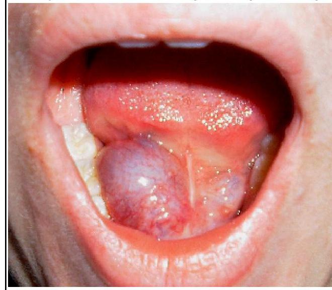
What is the most likely diagnosis for the swelling in the floor of the mouth shown in the image?
Q52
After laparoscopic cholecystectomy what should be the urine output of the patient if the renal function of the patient is normal?
Q53
What is the baseline platelet count required for surgery?