INI-CET 2023 — Biochemistry
9 Previous Year Questions with Answers & Explanations
Pompe disease is seen due to deficiency of?
Xanthurenic aciduria is seen in which vitamin deficiency?
BARTH syndrome is caused by deficiency of?
DNA sequence is determined by?
Liver produces ketones but cannot use it due to the deficiency of which of the following enzyme?
A urine sample shows a maroon-colored ring at the interface of two layers after adding reagents. Identify the test:
What is the most common cause of pure gonadal dysgenesis with XY karyotype?
Which of the following is used to detect abnormal gene sequences EXCEPT?
Which membrane channel is mainly affected in Cystic fibrosis?
INI-CET 2023 - Biochemistry INI-CET Practice Questions and MCQs
Question 1: Pompe disease is seen due to deficiency of?
- A. β-galactocerebrosidase
- B. β-glucocerebrosidase
- C. Lysosomal acid alpha glucosidase (Correct Answer)
- D. β-hexosaminidase-A
Explanation: ***Lysosomal acid alpha glucosidase*** - **Pompe disease**, also known as **glycogen storage disease type II**, is caused by the deficiency of **acid alpha-glucosidase (GAA)**, a lysosomal enzyme. - This deficiency leads to the accumulation of **glycogen** within lysosomes in various tissues, particularly muscle, liver, and heart, causing muscle weakness and cardiomegaly. *β-galactocerebrosidase* - Deficiency of **β-galactocerebrosidase** is associated with **Krabbe disease** (globoid cell leukodystrophy). - Krabbe disease involves the buildup of **galactocerebroside**, leading to progressive destruction of the myelin sheath in the central and peripheral nervous systems. *β-glucocerebrosidase* - Deficiency in **β-glucocerebrosidase** causes **Gaucher disease**, the most common lysosomal storage disorder. - This results in the accumulation of **glucocerebroside** in macrophages, leading to hepatosplenomegaly, bone pain, and neurologic symptoms in some forms. *β-hexosaminidase-A* - Deficiency of **β-hexosaminidase-A** is the underlying cause of **Tay-Sachs disease**. - Tay-Sachs disease is characterized by the accumulation of **GM2 gangliosides** in neurons, leading to progressive neurodegeneration, developmental delay, and a characteristic cherry-red spot in the retina.
Question 2: Xanthurenic aciduria is seen in which vitamin deficiency?
- A. Vitamin B5
- B. Vitamin B12
- C. Vitamin B6 (Correct Answer)
- D. Vitamin B7
Explanation: ***Vitamin B6*** - **Xanthurenic aciduria** is a classic sign of **Vitamin B6 (pyridoxine) deficiency** because B6 is a crucial coenzyme for **kynureninase**, an enzyme in the **tryptophan metabolic pathway**. - Without sufficient B6, tryptophan cannot be properly metabolized, leading to the accumulation and excretion of xanthurenic acid. *Vitamin B5* - **Vitamin B5 (pantothenic acid)** is a precursor to **coenzyme A (CoA)**, essential for fatty acid metabolism and the **Krebs cycle**. - Its deficiency is associated with symptoms like paresthesia and fatigue, not xanthurenic aciduria. *Vitamin B12* - **Vitamin B12 (cobalamin)** is critical for DNA synthesis and the formation of **red blood cells**, and its deficiency causes **megaloblastic anemia** and neurological symptoms. - It plays no direct role in the tryptophan-kynurenine pathway, so its deficiency does not lead to xanthurenic aciduria. *Vitamin B7* - **Vitamin B7 (biotin)** acts as a coenzyme in **carboxylase reactions**, involved in fatty acid synthesis, gluconeogenesis, and amino acid metabolism. - Deficiency can cause dermatitis, hair loss, and neurological issues, but it is not linked to xanthurenic acid accumulation.
Question 3: BARTH syndrome is caused by deficiency of?
- A. Glycolipids
- B. Sphingomyelin
- C. Cardiolipin (Correct Answer)
- D. Cerebroside
Explanation: ***Cardiolipin*** - **BARTH syndrome** is a rare, X-linked genetic disorder caused by mutations in the **TAZ gene**, which encodes for the enzyme **tafazzin**. - **Tafazzin** is crucial for the remodeling of **cardiolipin**, a phospholipid essential for mitochondrial membrane integrity and function. Deficiency of properly remodeled (mature) cardiolipin leads to the characteristic cardiomyopathy, skeletal myopathy, neutropenia, and growth delay seen in BARTH syndrome. *Glycolipids* - **Glycolipids** are lipids with a carbohydrate attached, important for cell recognition and signaling. - Their deficiency or abnormal metabolism is associated with conditions like **glycosphingolipidoses** (e.g., Gaucher disease, Fabry disease), not BARTH syndrome. *Sphingomyelin* - **Sphingomyelin** is a type of sphingolipid found in animal cell membranes, particularly in the myelin sheath. - Its deficiency or accumulation is linked to **Niemann-Pick disease**, which presents with hepatosplenomegaly and neurodegeneration, distinct from BARTH syndrome. *Cerebroside* - **Cerebrosides** are a type of glycosphingolipid found in the myelin sheath and nerve cell membranes. - Disorders involving cerebroside metabolism include **Krabbe disease** (globoid cell leukodystrophy) and **Gaucher disease**, which are pathologically distinct from BARTH syndrome.
Question 4: DNA sequence is determined by?
- A. Sanger sequencing (Correct Answer)
- B. PCR
- C. FISH
- D. Gel electrophoresis
Explanation: ***Correct: Sanger sequencing*** - **Sanger sequencing** (chain-termination method) is the gold standard technique used to determine the exact order of nucleotides within a DNA molecule - It uses dideoxynucleotides (ddNTPs) to terminate DNA strand elongation at specific bases, producing fragments of varying lengths - These fragments are separated by capillary electrophoresis and the sequence is read based on the terminal fluorescent label - Directly determines DNA sequence with high accuracy *Incorrect: PCR* - **Polymerase Chain Reaction (PCR)** amplifies specific DNA segments to create millions of copies - It does NOT determine the sequence itself - it only makes copies of DNA - PCR-amplified DNA can be used as a template for subsequent sequencing, but PCR itself doesn't reveal sequence information *Incorrect: FISH* - **Fluorescence in situ hybridization (FISH)** detects and localizes specific DNA sequences on chromosomes - Used for chromosomal mapping and detecting chromosomal abnormalities - Does not determine the nucleotide sequence *Incorrect: Gel electrophoresis* - Separates DNA fragments based on size and charge - Used to analyze DNA but cannot determine the specific nucleotide sequence - Useful for visualizing DNA after amplification or restriction digestion
Question 5: Liver produces ketones but cannot use it due to the deficiency of which of the following enzyme?
- A. Alkaline phosphatase
- B. Alanine transaminase
- C. Thiophorase (Correct Answer)
- D. Thiolase
Explanation: ***Thiophorase*** - The liver lacks **thiophorase (succinyl-CoA:3-ketoacid CoA transferase)**, which is crucial for converting **acetoacetate** to **acetoacetyl-CoA**. - This enzyme deficiency prevents the liver from utilizing ketones as an energy source, even though it is a primary site for their production. *Alkaline phosphatase* - **Alkaline phosphatase** is a non-specific enzyme found in various tissues, including bone, liver, and intestine. - Its primary role is to **hydrolyze phosphate esters**, and it is not directly involved in ketone metabolism. *Alanine transaminase* - **Alanine transaminase (ALT)** is a liver enzyme primarily involved in **amino acid metabolism**, specifically in the transfer of an amino group from alanine to α-ketoglutarate. - It plays no direct role in the synthesis or utilization of ketone bodies. *Thiolase* - **Thiolase** is an enzyme involved in both the synthesis and breakdown of ketone bodies. - It converts **two acetyl-CoA molecules into acetoacetyl-CoA** during ketogenesis and also cleaves acetoacetyl-CoA into two acetyl-CoA molecules during ketolysis in extrahepatic tissues.
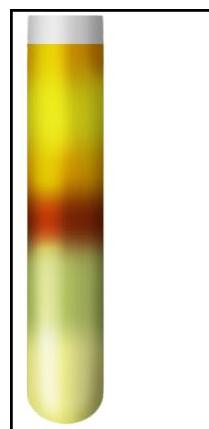
Question 6: A urine sample shows a maroon-colored ring at the interface of two layers after adding reagents. Identify the test:
- A. Rothera's test (Correct Answer)
- B. Benedict's test
- C. Biuret test
- D. Fehling's test
Explanation: ***Rothera's test*** - The image displays a **maroon-colored ring** at the interface of two layers, which is a characteristic positive result for Rothera's test. - Rothera's test is used to detect the presence of **ketone bodies**, specifically **acetone and acetoacetate**, in urine. *Benedict's test* - Benedict's test is used to detect **reducing sugars**, such as glucose, in urine. - A positive Benedict's test typically produces a color change from blue to green, yellow, orange, or brick-red precipitate after heating, which is not what is seen here. *Biuret test* - The Biuret test is used to detect **peptide bonds** and thus the presence of **proteins**. - A positive Biuret test results in a **violet or purple color** when proteins are present, which is different from the reaction shown. *Fehling's test* - Fehling's test is another test used to detect the presence of **reducing sugars** in a sample. - A positive result is indicated by the formation of a **brick-red precipitate** of cuprous oxide after heating, which is not consistent with the image.
Question 7: What is the most common cause of pure gonadal dysgenesis with XY karyotype?
- A. Deletion
- B. Point mutation (Correct Answer)
- C. Insertion
- D. Translocation
Explanation: Point mutation - The most common genetic cause of **pure gonadal dysgenesis with XY karyotype** (Swyer syndrome) is a **point mutation** within the **SRY gene**, located on the Y chromosome [1]. - Point mutations include **missense mutations** (amino acid substitution) and **nonsense mutations** (premature stop codon), both of which can render the SRY protein non-functional. - This specific type of mutation leads to a non-functional SRY protein, preventing the development of testes in an individual with an XY karyotype and resulting in streak gonads [1]. *Deletion* - While deletions involving the SRY gene can cause XY gonadal dysgenesis, **complete deletion** of SRY is less common than point mutations as the primary cause of pure gonadal dysgenesis. - Larger deletions on the Y chromosome might also affect other genes, leading to a broader spectrum of phenotypes. *Insertion* - **Insertions** are a type of frameshift mutation where nucleotides are added to the DNA sequence. - While insertions in the SRY gene could theoretically cause gonadal dysgenesis, they are much less commonly reported than point mutations as the cause of Swyer syndrome. *Translocation* - **Translocations** involving the SRY gene, such as SRY being translocated to an X chromosome, can lead to sex reversal (XX male phenotype). - However, for pure XY gonadal dysgenesis (where SRY is present but non-functional), translocations are not the most common underlying genetic mechanism.
Question 8: Which of the following is used to detect abnormal gene sequences EXCEPT?
- A. RFLP analysis
- B. Pyrosequencing
- C. Flow cytometry (Correct Answer)
- D. FISH
Explanation: ***Flow cytometry*** - **Flow cytometry** is primarily used to analyze **cell populations** based on their physical and biochemical characteristics (e.g., size, granularity, and protein expression) by passing them single file through a laser beam, not for direct gene sequencing. - It detects and quantifies cells labeled with **fluorescent antibodies**, making it useful for immunophenotyping, cell sorting, and DNA content analysis, but not for identifying specific gene sequences or mutations. *RFLP analysis* - **Restriction fragment length polymorphism (RFLP) analysis** detects variations in **DNA sequences** by using **restriction enzymes** to cut DNA at specific sites. - Differences in fragment lengths indicate **polymorphisms** or **mutations** within the recognition sites, thereby identifying abnormal gene sequences. *Pyrosequencing* - **Pyrosequencing** is a method of **DNA sequencing** that determines the sequence of nucleotides by detecting the release of pyrophosphate during DNA synthesis. - It is used to identify **single nucleotide polymorphisms (SNPs)** and **short genetic variations**, making it suitable for detecting abnormal gene sequences. *FISH* - **Fluorescence in situ hybridization (FISH)** uses **fluorescently labeled DNA probes** that bind to specific complementary **DNA sequences** on chromosomes. - It is a powerful cytogenetic technique for detecting **chromosomal abnormalities**, such as deletions, translocations, and amplifications, thereby identifying abnormal gene sequences.
Question 9: Which membrane channel is mainly affected in Cystic fibrosis?
- A. Sodium
- B. Chloride (Correct Answer)
- C. Calcium
- D. Potassium
Explanation: ***Chloride*** - Cystic fibrosis is caused by a mutation in the **CFTR (Cystic Fibrosis Transmembrane Conductance Regulator)** gene, which encodes a chloride channel. - Dysfunction of this **chloride channel** leads to impaired transport of chloride ions, mainly affecting epithelial cells in various organs. *Sodium* - While sodium transport is indirectly affected in cystic fibrosis, the primary defect is not in a sodium channel itself but rather in the **chloride channel**, which influences water and sodium movement. - The abnormal **chloride transport** leads to a compensatory but ineffective increase in sodium absorption in some tissues like the airway. *Calcium* - Calcium channels are not primarily implicated in the pathophysiology of **cystic fibrosis**. - **Calcium dysregulation** can occur secondarily in some CF-related processes, but it is not the main affected membrane channel. *Potassium* - **Potassium channels** are not the main membrane channels affected in cystic fibrosis. - While potassium transport is vital for cellular function, it is not the primary defect underlying the disease's respiratory and gastrointestinal manifestations.