INI-CET 2023 — Anatomy
9 Previous Year Questions with Answers & Explanations
What can cause the absence of fructose in seminal fluid?
Identify the position of the appendix marked in BLACK in the given image:
What is the incorrect statement?
What is the correct sequence of the auditory pathway?
Identify the lines shown in the following image:
Which of the following structures is supplied by the superior gluteal nerve?
What is the action of the muscle shown in the image below?
Match the following: A) Glossopharyngeal nerve B) Spinal accessory nerve C) Facial nerve D) Mandibular nerve 1) Shrugging of shoulder 2) Touch sensation from the posterior one-third of the tongue 3) Chewing 4) Taste from the anterior two-thirds of the tongue
Anterior relations of third part of duodenum are all except?
INI-CET 2023 - Anatomy INI-CET Practice Questions and MCQs
Question 1: What can cause the absence of fructose in seminal fluid?
- A. None of the above
- B. Congenital absence of seminal vesicle
- C. Ejaculatory duct obstruction
- D. Both of the above (Correct Answer)
Explanation: ***Both of the above*** - **Fructose** in seminal fluid is primarily produced by the **seminal vesicles**, providing energy for sperm motility. - Therefore, either a **congenital absence of seminal vesicles** or an **ejaculatory duct obstruction** (preventing seminal vesicle secretions from reaching the ejaculate) would lead to the absence of fructose. *Congenital absence of seminal vesicle* - The **seminal vesicles** are the primary source of fructose in seminal fluid. - If a person is born without these glands, **fructose will be absent** from their seminal fluid. *Ejaculatory duct obstruction* - An obstruction in the **ejaculatory ducts** would block the passage of secretions from the **seminal vesicles** and vasoepididymis into the urethra. - This prevents **fructose** (from the seminal vesicles) and sperm (from the testes/epididymis) from being present in the ejaculate. *None of the above* - This option is incorrect because both **congenital absence of seminal vesicles** and **ejaculatory duct obstruction** are valid causes for the absence of fructose in seminal fluid.
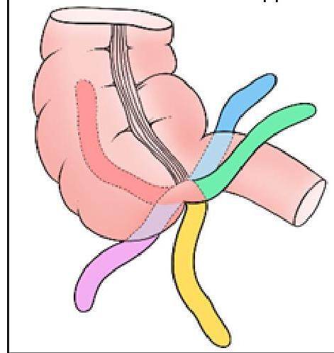
Question 2: Identify the position of the appendix marked in BLACK in the given image:
- A. Pelvic
- B. Subcecal
- C. Retrocecal (Correct Answer)
- D. Preileal
Explanation: ***Retrocecal*** - The **retrocecal** position (represented by the black color in the image) indicates the appendix is located behind the cecum, often a common variant. - This position can make diagnosis of appendicitis challenging as it may cause atypical pain patterns. *Pelvic* - The **pelvic** appendix descends into the true pelvis, which can mimic gynecological or urological conditions. - It usually causes pain that is more generalized in the lower abdomen or suprapubic region. *Subcecal* - The **subcecal** appendix is located directly below the cecum and is a relatively rare position. - While somewhat straightforward in presentation, it is less common than retrocecal or pelvic positions. *Preileal* - The **preileal** position indicates the appendix lies in front of the terminal ileum. - This is a less common anatomical variation, often associated with specific clinical presentations related to its anterior location.
Question 3: What is the incorrect statement?
- A. MIS inhibits the formation of Mullerian duct
- B. WD form male internal genitalia
- C. Zygote is Bipotential at 8 weeks (Correct Answer)
- D. DHT is necessary for the development of external genitals
Explanation: ***Zygote is Bipotential at 8 weeks*** - A **zygote** is formed at conception and is the single-cell diploid organism, not bipotential at 8 weeks. - The **bipotential gonad** can develop into either testes or ovaries, and this stage of sexual differentiation occurs earlier in gestation, typically around the 6th to 7th week, before differentiating into male or female gonads, not at 8 weeks as an entire zygote. *MIS inhibits the formation of Mullerian duct* - **Müllerian Inhibiting Substance (MIS)**, also known as **Anti-Müllerian Hormone (AMH)**, is produced by the Sertoli cells of the developing testes [1]. - Its primary function is to cause the **regression of the Müllerian ducts**, which would otherwise develop into female internal reproductive structures (fallopian tubes, uterus, and upper vagina) [1]. *WD form male internal genitalia* - The **Wolffian ducts (WD)**, also known as mesonephric ducts, are precursors to male internal genitalia in the presence of testosterone [1]. - stimulated by **testosterone** produced by the Leydig cells of the fetal testes, they develop into the **epididymis, vas deferens, and seminal vesicles** [1]. *DHT is necessary for the development of external genitals* - **Dihydrotestosterone (DHT)**, a more potent form of testosterone, is crucial for the development of male external genitalia [1]. - The enzyme **5α-reductase** converts testosterone to DHT in target tissues, leading to the formation of the **penis, scrotum, and prostate** [1].
Question 4: What is the correct sequence of the auditory pathway?
- A. Spiral Ganglion → Cochlea → Cochlear Nerve → Superior Olivary N
- B. Spiral Ganglion → Cochlear Nerve → Cochlea → Superior Olivary N
- C. Cochlea → Spiral Ganglion → Cochlear Nerve → Superior Olivary N (Correct Answer)
- D. Cochlear Nerve → Spiral Ganglion → Cochlea → Superior Olivary N
Explanation: ***Cochlea → Spiral Ganglion → Cochlear Nerve → Superior Olivary N*** - Sound vibrations are first transduced into electrical signals by the **hair cells** in the **cochlea** [2]. These signals are then transmitted to the **spiral ganglion**. - Neurons in the **spiral ganglion** generate action potentials, which are carried by the **cochlear nerve** to the brainstem, specifically the **superior olivary nucleus**, for further processing [1]. *Spiral Ganglion → Cochlea → Cochlear Nerve → Superior Olivary N* - This sequence is incorrect because the **cochlea** is where the initial mechanical-to-electrical transduction of sound occurs, *before* the signal reaches the **spiral ganglion** neurons [2]. - The spiral ganglion consists of the cell bodies of the neurons that innervate the cochlea's hair cells, meaning the cochlea must process the sound first. *Spiral Ganglion → Cochlear Nerve → Cochlea → Superior Olivary N* - This order is incorrect as the **cochlea** is the organ that processes sound input *prior* to the involvement of the **spiral ganglion** and the **cochlear nerve** [2]. - The flow of information begins at the peripheral sensory organ (cochlea) and then moves centrally. *Cochlear Nerve → Spiral Ganglion → Cochlea → Superior Olivary N* - This sequence is incorrect because the **cochlea** is the initial site of sound detection and signal generation, *before* the **cochlear nerve** transmits the signal. - The **spiral ganglion** contains the cell bodies of the neurons whose axons form the cochlear nerve, so the signal must pass through the ganglion before going down the nerve.
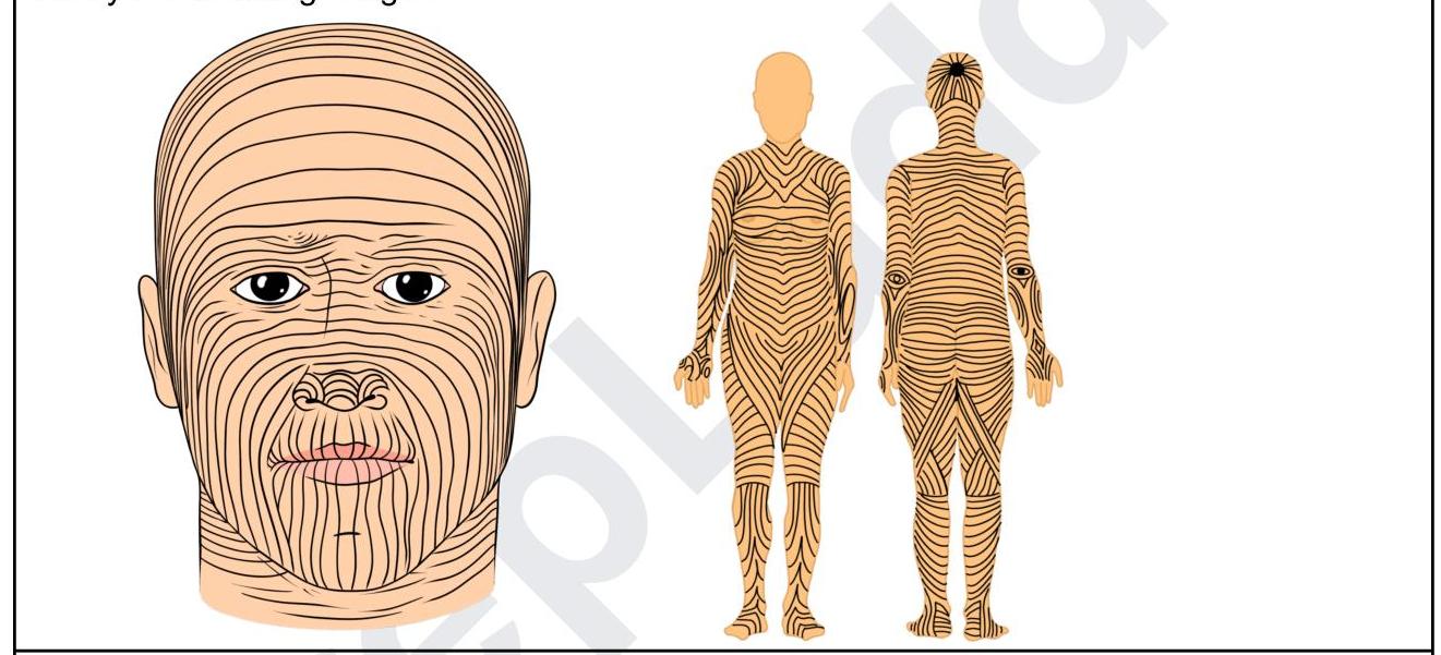
Question 5: Identify the lines shown in the following image:
- A. Hinderer's lines
- B. Dermatomes
- C. Langer's lines (Correct Answer)
- D. Blaschko's lines
Explanation: ***Langer's lines*** - The image displays lines that represent the **natural orientation of collagen fibers** within the human skin, which are known as Langer's lines (also called cleavage lines). - Making surgical incisions **parallel to these lines** can result in better wound healing and less scarring. - Named after **Karl Langer**, an Austrian anatomist who described these lines in 1861. *Hinderer's lines* - While **Hinderer** described relaxed skin tension lines (RSTLs) used in plastic surgery, these are **different from Langer's lines**. - The image shows Langer's lines specifically, which are based on **collagen fiber orientation**, not relaxed skin tension. *Dermatomes* - **Dermatomes** are areas of skin mainly supplied by a single **spinal nerve root**. - They represent **neurologic segments** and do not correspond to the collagen fiber orientation shown in the image. *Blaschko's lines* - **Blaschko's lines** are invisible lines of skin cell migration that become visible in certain **genetic or acquired dermatological conditions**. - They represent a **mosaic pattern** due to different cell populations and are distinctly different from the structural collagen lines shown.
Question 6: Which of the following structures is supplied by the superior gluteal nerve?
- A. Gluteus minimus (Correct Answer)
- B. Gluteus maximus
- C. Piriformis
- D. All of the options
Explanation: ***Gluteus minimus*** - The **superior gluteal nerve** provides motor innervation to the gluteus medius, gluteus minimus, and tensor fasciae latae muscles. - This nerve originates from the sacral plexus **(L4, L5, S1)** and exits the pelvis through the greater sciatic foramen, superior to the piriformis muscle. *Gluteus maximus* - The gluteus maximus muscle is innervated by the **inferior gluteal nerve**, not the superior gluteal nerve. - The inferior gluteal nerve also arises from the sacral plexus **(L5, S1, S2)** and is crucial for hip extension and external rotation. *Piriformis* - The piriformis muscle receives its own direct branches from the sacral plexus **(S1, S2)** via the nerve to piriformis, distinct from the superior or inferior gluteal nerves. - It plays a key role in hip external rotation and abduction when the hip is flexed. *All of the options* - This option is incorrect because gluteus maximus is innervated by the inferior gluteal nerve, and piriformis has its own specific nerve supply. - The superior gluteal nerve specifically innervates only the gluteus medius, gluteus minimus, and tensor fasciae latae.
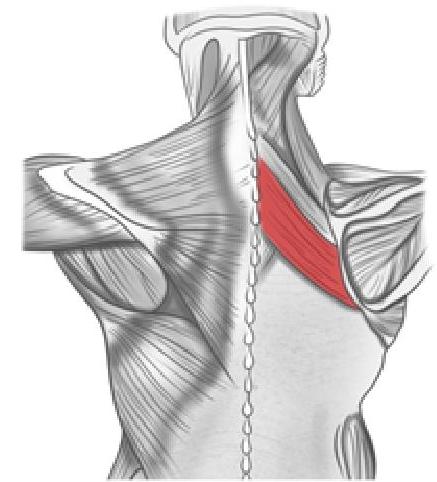
Question 7: What is the action of the muscle shown in the image below?
- A. Medial rotation of the shoulder
- B. Adduction of the shoulder
- C. Extension of the shoulder
- D. Retracts the scapula (Correct Answer)
Explanation: ***Retracts the scapula*** - The image highlights the **rhomboid major** muscle, which originates from the spinous processes of T2-T5 vertebrae and inserts onto the medial border of the scapula. - Its primary action is to **retract** (pull medially) and rotate the scapula inferiorly, and also to help hold the scapula against the thoracic wall. *Medial rotation of the shoulder* - Medial rotation of the shoulder is primarily performed by muscles like the **subscapularis**, **pectoralis major**, **latissimus dorsi**, and **teres major**. - The rhomboids do not directly act on the glenohumeral joint for shoulder rotation. *Adduction of the shoulder* - Adduction of the shoulder (bringing the arm towards the body) is mainly performed by the **latissimus dorsi**, **pectoralis major**, and **teres major**. - The rhomboids' action on the scapula indirectly influences shoulder movement but does not directly adduct the shoulder joint. *Extension of the shoulder* - Shoulder extension is primarily achieved by the **latissimus dorsi**, **teres major**, and the posterior fibers of the **deltoid**. - The rhomboid major muscle's action focuses solely on the scapula, not direct extension of the shoulder joint.
Question 8: Match the following: A) Glossopharyngeal nerve B) Spinal accessory nerve C) Facial nerve D) Mandibular nerve 1) Shrugging of shoulder 2) Touch sensation from the posterior one-third of the tongue 3) Chewing 4) Taste from the anterior two-thirds of the tongue
- A. A-3 , B-1 , C-4 , D-2
- B. A-2 , B-3 , C-4 , D-1
- C. A-4 , B-1 , C-2 , D-3
- D. A-2 , B-1 , C-4 , D-3 (Correct Answer)
Explanation: ***A-2 , B-1 , C-4 , D-3*** - **A) Glossopharyngeal nerve (CN IX)** is responsible for **general sensation and taste from the posterior one-third of the tongue** [1]. (2). - **B) Spinal Accessory nerve (CN XI)** innervates the **sternocleidomastoid** and **trapezius muscles**, which are involved in shrugging the shoulders (1). - **C) Facial nerve (CN VII)** carries **taste sensation from the anterior two-thirds of the tongue** [1] (4) via the chorda tympani. - **D) Mandibular nerve (V3)**, a branch of the trigeminal nerve, innervates the muscles of mastication, enabling **chewing** (3). *A-3 , B-1 , C-4 , D-2* - This option incorrectly associates the **glossopharyngeal nerve** with chewing, which is a function of the mandibular nerve (V3). - It also incorrectly associates the **mandibular nerve** with touch sensation from the posterior one-third of the tongue, which is a function of the glossopharyngeal nerve [1]. *A-2 , B-3 , C-4 , D-1* - This option incorrectly links the **spinal accessory nerve** with chewing; this nerve primarily controls shoulder and neck movements. - It also incorrectly assigns shrugging of the shoulder to the **mandibular nerve** instead of the spinal accessory nerve. *A-4 , B-1 , C-2 , D-3* - This choice incorrectly attributes **taste from the anterior two-thirds of the tongue** to the glossopharyngeal nerve, which supplies the posterior one-third [1]. - It also incorrectly links **touch sensation from the posterior one-third of the tongue** to the facial nerve, which is involved in taste from the anterior two-thirds [1].
Question 9: Anterior relations of third part of duodenum are all except?
- A. Jejunum
- B. Fundus of gallbladder (Correct Answer)
- C. Root of mesentery
- D. Superior mesenteric artery
Explanation: ***Fundus of gallbladder*** - The **fundus of the gallbladder** is located more superiorly and anteriorly, typically lying near the ninth costal cartilage, and is not an anterior relation of the third part of the duodenum. - The third part of the duodenum lies mainly at the level of the **L3 vertebra**, far removed from the gallbladder fundus. *Jejunum* - The **jejunum**, being part of the mobile small intestine, can lie anterior to the third part of the duodenum. - These two structures are anatomically close and can overlap. *Root of mesentery* - The **root of the mesentery** crosses anterior to the third part of the duodenum, attaching to the posterior abdominal wall. - This is a key anatomical landmark that helps fix the position of the small intestine. *Superior mesenteric artery* - The **superior mesenteric artery** and vein both cross **anterior** to the third part of the duodenum as they emerge from beneath the pancreas. - This anatomical relationship is clinically relevant in conditions like superior mesenteric artery syndrome.