INI-CET 2022 — Radiology
4 Previous Year Questions with Answers & Explanations
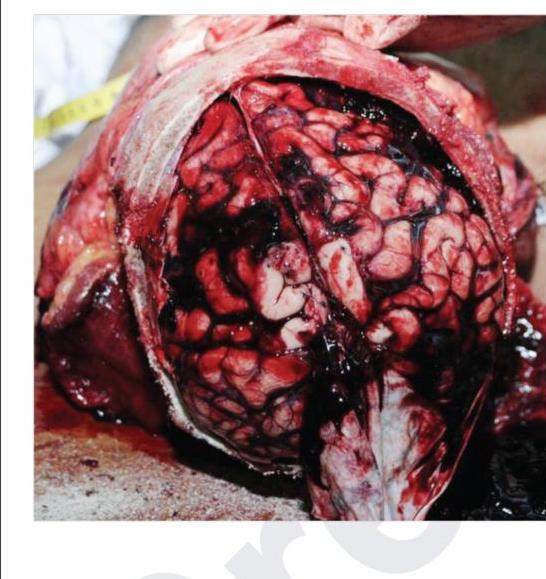
Bleeding as shown in the image is due to which of the following vessels?
Which of the following findings are seen in a high-resolution CT scan of fungal pneumonia? 1. Interlobular septations 2. Peripheral wedge-shaped consolidation 3. Pleural effusion 4. Cavitatory lesions with surrounding ground glass opacities
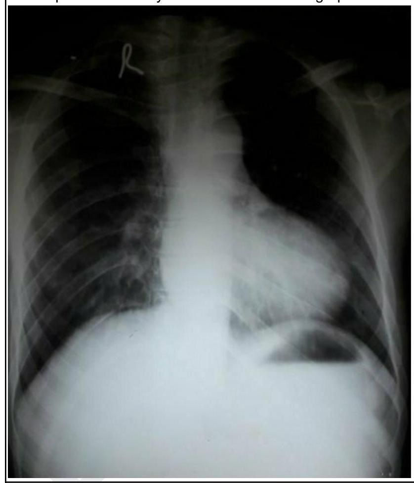
A child presents with cyanosis. His chest radiograph is shown below. What is the diagnosis?
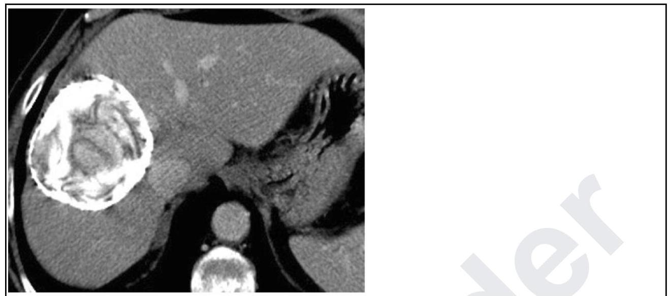
Identify the condition based on the non-contrast CT scan of a patient given below.
INI-CET 2022 - Radiology INI-CET Practice Questions and MCQs
Question 1: Bleeding as shown in the image is due to which of the following vessels?
- A. Lenticulostriate artery
- B. Vertebral artery
- C. Bridging veins (Correct Answer)
- D. Middle meningeal artery
Explanation: ***Bridging veins*** - The image depicts a **subdural hemorrhage (subdural hematoma)**, a collection of blood between the dura mater and the arachnoid mater, typically appearing as a **crescent-shaped** hyperdensity that conforms to the brain surface. - This type of hemorrhage is caused by the tearing of **bridging veins** that traverse the subdural space, connecting the cerebral cortex to the dural venous sinuses. - Tearing of these veins occurs due to rapid acceleration-deceleration forces causing the brain to move relative to the dura, stretching and rupturing the veins. This is common in **head trauma**, especially in the elderly (due to brain atrophy increasing vein vulnerability) or infants. *Lenticulostriate artery* - Rupture of the lenticulostriate arteries (perforating branches of the middle cerebral artery) typically leads to **intraparenchymal hemorrhage**, specifically in the basal ganglia or internal capsule. - This type of bleeding is confined within the brain parenchyma, rather than collecting in the subdural space as seen in the image. - Associated with hypertensive hemorrhage. *Vertebral artery* - The vertebral arteries supply the posterior circulation of the brain, and their rupture can lead to **subarachnoid hemorrhage** (if a posterior circulation aneurysm ruptures) or **intraparenchymal bleeding** in the brainstem or cerebellum. - Bleeding from the vertebral artery is not associated with the subdural collection pattern shown in the image. *Middle meningeal artery* - The middle meningeal artery runs in the epidural space, and its rupture (often due to temporal bone fracture) causes an **epidural hematoma**. - An epidural hematoma is characterized by a **biconvex (lentiform) shape** on imaging and is situated between the dura mater and the skull, which is distinct from the **crescent-shaped** subdural collection shown. - Does not cross suture lines, unlike subdural hematomas which can extend over multiple lobes.
Question 2: Which of the following findings are seen in a high-resolution CT scan of fungal pneumonia? 1. Interlobular septations 2. Peripheral wedge-shaped consolidation 3. Pleural effusion 4. Cavitatory lesions with surrounding ground glass opacities
- A. 1,2,3
- B. 2,3,4
- C. 1,2,4 (Correct Answer)
- D. 1,3,4
Explanation: ***1,2,4*** - **Interlobular septations** and **peripheral wedge-shaped consolidations** are common findings due to the **vascular invasion** and **infarction** characteristic of fungal pneumonia. - **Cavitary lesions with surrounding ground-glass opacity**, also known as the **halo sign**, are highly suggestive of invasive fungal infections like aspergillosis. *1,2,3* - While interlobular septations and peripheral wedge-shaped consolidations are seen in fungal pneumonia, **pleural effusion** is less common and not a primary diagnostic feature. - The absence of the characteristic cavitary lesions with ground-glass opacities makes this option incomplete. *2,3,4* - This option correctly includes peripheral wedge-shaped consolidation and cavitary lesions with ground-glass opacity, but the inclusion of **pleural effusion** and exclusion of **interlobular septations** make it less accurate. - Interlobular septations are a significant indicator of **lymphatic involvement** as seen in fungal diseases. *1,3,4* - Although interlobular septations and cavitary lesions with ground-glass opacities are relevant, the presence of **pleural effusion** as a primary finding is less typical for fungal pneumonia. - The absence of **peripheral wedge-shaped consolidation**, which arises from vascular occlusion, makes this option less comprehensive.
Question 3: A child presents with cyanosis. His chest radiograph is shown below. What is the diagnosis?
- A. Transposition of great arteries (TGA)
- B. Ebstein's anomaly
- C. Tetralogy of Fallot (TOF) (Correct Answer)
- D. Total anomalous pulmonary venous return (TAPVC)
Explanation: ***Tetralogy of Fallot (TOF)*** - The chest radiograph shows a **boot-shaped heart (coeur en sabot)** due to **right ventricular hypertrophy** and a **concave pulmonary artery segment** (absent main pulmonary artery segment), which is characteristic of TOF. - The patient also presents with **cyanosis**, a common symptom of TOF due to right-to-left shunting. *Transposition of great arteries (TGA)* - TGA typically presents with a **"egg-on-a-string" appearance** on chest radiograph, indicating a narrow superior mediastinum and increased pulmonary vascular markings, which is not seen here. - While patients with TGA are cyanotic, the cardiac silhouette on this radiograph is inconsistent with the classic TGA findings. *Ebstein's anomaly* - Ebstein's anomaly is characterized by **apical displacement of the tricuspid valve**, leading to a large right atrium and massive **cardiomegaly** on chest X-ray, often described as a "box-shaped" heart, which is not evident in the provided image. - While it causes cyanosis, the heart size in the image is not markedly enlarged enough to suggest Ebstein's anomaly. *Total anomalous pulmonary venous return (TAPVC)* - TAPVC typically presents with a **"snowman" or "figure-of-8" appearance** on chest X-ray due to a dilated superior vena cava and left brachiocephalic vein, or a small heart with increased pulmonary vascularity, neither of which is present in the image. - Although TAPVC causes cyanosis, the specific radiographic features like the "boot-shaped" heart rule out this diagnosis.
Question 4: Identify the condition based on the non-contrast CT scan of a patient given below.
- A. Hepatocellular carcinoma
- B. Hydatid cyst (Correct Answer)
- C. Liver abscess
- D. Focal nodular hyperplasia
Explanation: ***Hydatid cyst*** - The image distinctly shows a **large, well-defined cyst with internal septations**, consistent with the daughter cysts and collapsed membranes within a hydatid cyst (the "**water lily sign**"). - The thick, often calcified wall surrounding the lesion is a characteristic feature often seen in **Echinococcus granulosa** infection. *Hepatocellular carcinoma* - **Hepatocellular carcinoma (HCC)** typically appears as a **solid, enhancing mass** (especially on contrast-enhanced CT) and does not usually present with clearly defined internal septations or "water lily" sign on non-contrast imaging. - While HCC can show necrosis, it does not form the characteristic cystic structure seen here. *Liver abscess* - A **liver abscess** would typically appear as a ill-defined, fluid-filled lesion that may have a rim of enhancement on contrast CT, but it generally lacks the **distinct internal septations** or daughter cysts characteristic of a hydatid cyst. - Abscesses are often associated with signs of infection like fever and elevated inflammatory markers. *Fibronodular hyperplasia* - **Focal nodular hyperplasia (FNH)** is a benign liver lesion characterized by a central scar and is typically **isodense or slightly hypodense** to the liver parenchyma on non-contrast CT. - It does not present as a cystic lesion with internal daughter cysts or calcified walls.