All (164)Anatomy (15)Anesthesiology (1)Biochemistry (8)Community Medicine (11)Dermatology (7)ENT (2)Forensic Medicine (7)Internal Medicine (16)Microbiology (13)Obstetrics and Gynecology (9)Ophthalmology (3)Orthopaedics (2)Pathology (16)Pediatrics (8)Pharmacology (14)Physiology (11)Psychiatry (4)Radiology (5)Surgery (12)
Q31
Deamination of methylated cytosine forms which of the following?
Q32
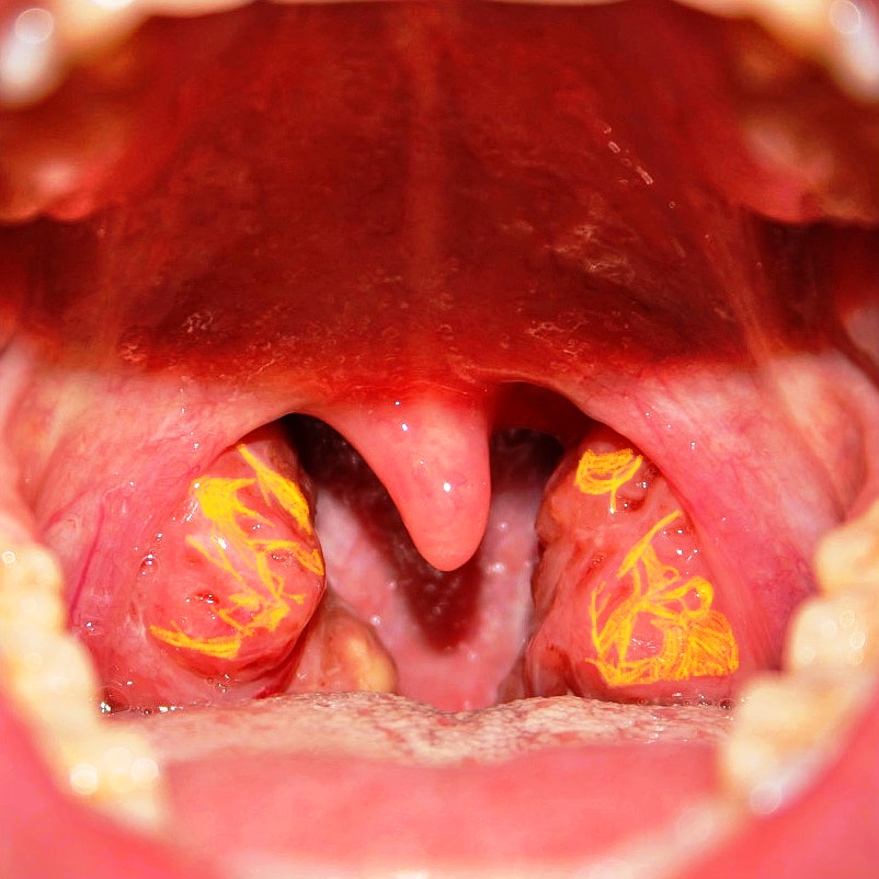
A patient presented with reduced levels of high-density lipoprotein, and ABCA1 mutation. On examination, tonsils appeared as shown in the image. What is the diagnosis?
Q33
Increased H+ ions in the intermembrane space of mitochondria are due to?