All (164)Anatomy (15)Anesthesiology (1)Biochemistry (8)Community Medicine (11)Dermatology (7)ENT (2)Forensic Medicine (7)Internal Medicine (16)Microbiology (13)Obstetrics and Gynecology (9)Ophthalmology (3)Orthopaedics (2)Pathology (16)Pediatrics (8)Pharmacology (14)Physiology (11)Psychiatry (4)Radiology (5)Surgery (12)
Q21
Which ligament connects medial cuneiform to the base of the 2nd metatarsal?
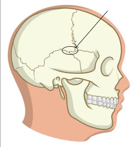
Q22
Which structure is supplied by the nerve causing this elevation?
Q23
Arrange lung hilar structure from anterior to posterior:- 1. Primary bronchus 2. Bronchial artery 3. Pulmonary vein 4. Pulmonary artery
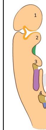
Q24
A child lacks thymus and inferior parathyroid glands. Defective development of which of the following structures is likely to be the cause?
Q25
Arrange the cells according to their positions from the basal layer towards the lumen in the seminiferous tubules:- 1. Spermatogonia 2. Primary spermatocyte 3. Spermatid 4. Spermatozoa
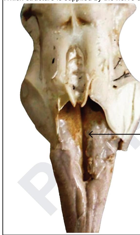
Q26
Identify the incorrect statement regarding the marked structure.