INI-CET 2022 — Microbiology
11 Previous Year Questions with Answers & Explanations
Affinity maturation of antibodies is because of _____ .
Antigen presented on MHC class I molecules activates which of the following cells?
Which of the following is true about anti-CMV IgG antibodies?
Which of the following statements regarding the given image is correct?
Which of the following are acid-fast staining organisms? 1. Nocardia 2. Mycobacterium leprae 3. Actinomyces 4. Cryptosporidium parvum 5. Isospora belli
A young man presents with skin lesions as shown in the image below. All of the following organisms can spread through dermal and subcutaneous lymphatics, except
Which virus can be identified by a PCR method and is endemic to India?
Which of the following is a gram-positive organism that shows the following appearance on Ziehl-Neelsen staining?
A forest worker developed skin lesions over the forearm, which initially started as macules but then became nodules. Histology of the nodule shows the following findings. Which of the following is true regarding this condition?
The viruses of the Filoviridae family like Ebola and Marburg resemble which of the following morphologies?
INI-CET 2022 - Microbiology INI-CET Practice Questions and MCQs
Question 1: Affinity maturation of antibodies is because of _____ .
- A. Gene rearrangements
- B. CD40
- C. Somatic hypermutation (Correct Answer)
- D. Differential mRNA processing
Explanation: ***Somatic hypermutation*** - **Somatic hypermutation** is a process that introduces point mutations in the **variable regions** of immunoglobulin genes, primarily in B cells. - These mutations lead to the production of B cells with slightly altered **antibody affinities**, allowing for selection of those with higher affinity for the antigen. *Gene rearrangements* - **Gene rearrangements**, specifically **V(D)J recombination**, are responsible for the initial diversity of antibody specificities in immature B cells. - This process determines the basic antigen-binding site but does not fine-tune the **affinity** after initial antigen exposure. *CD40* - **CD40** is a co-stimulatory molecule on B cells that binds to **CD40L** on T cells, crucial for B cell activation, **isotype switching**, and germinal center formation. - While essential for antibody responses and germinal center reactions where affinity maturation occurs, **CD40** itself does not directly cause the molecular changes that lead to affinity maturation. *Differential mRNA processing* - **Differential mRNA processing** (or alternative splicing) primarily controls the production of different protein isoforms from a single gene. - In the context of antibodies, it can determine whether a B cell produces **membrane-bound** or **secreted** forms of antibodies, but it does not enhance the antigen-binding affinity.
Question 2: Antigen presented on MHC class I molecules activates which of the following cells?
- A. NK cells
- B. Helper cells
- C. B cells
- D. Cytotoxic T cells (Correct Answer)
Explanation: ***Cytotoxic T cells*** - **MHC class I** molecules present **intracellular antigens** (e.g., viral or tumor antigens) to **CD8+ T cells** (cytotoxic T cells). - This binding activates the cytotoxic T cells, leading to the destruction of the **infected** or **abnormal host cell**. *NK cells* - **Natural Killer (NK) cells** recognize and kill target cells that have **reduced or absent MHC class I** expression, which often occurs in virally infected or tumor cells, not cells presenting antigens on MHC class I. - They are part of the **innate immune system** and do not require prior sensitization or MHC-peptide presentation for activation. *Helper cells* - **Helper T cells** (CD4+ T cells) recognize antigens presented on **MHC class II** molecules, typically expressed by **antigen-presenting cells** (APCs) like macrophages, B cells, and dendritic cells. - Their primary role is to **orchestrate immune responses** by releasing cytokines. *B cells* - **B cells** are primarily involved in **humoral immunity**, producing antibodies after recognizing specific antigens directly via their B cell receptors or with T cell help. - While they can present antigens on **MHC class II** to helper T cells, direct antigen binding to **MHC class I** does not activate B cells.
Question 3: Which of the following is true about anti-CMV IgG antibodies?
- A. IgG avidity assay helps in differentiating past and primary infection (Correct Answer)
- B. Denotes latent CMV infection
- C. Denotes chronic CMV infection with immunity to other serotypes
- D. Indicates acute CMV infection
Explanation: ***IgG avidity assay helps in differentiating past and primary infection*** - **IgG avidity** measures the binding strength of IgG antibodies to their antigen. In a **primary infection**, IgG antibodies have low avidity. - As the immune response matures over several months, the avidity of IgG antibodies increases, indicating a **past infection**. *Denotes latent CMV infection* - While the presence of IgG antibodies indicates a past exposure and often a latent infection, it doesn't solely *denote* latency, as primary infection also involves IgG production. - **Latent CMV infection** specifically refers to the persistence of the virus in cells without active replication, which is usually confirmed by the presence of IgG antibodies but needs further contextual information like negative IgM and viral load. *Denotes chronic CMV infection with immunity to other serotypes* - CMV typically exists as one serotype, and IgG antibodies confer protection against *re-activation* of that specific virus, not immunity to "other serotypes." - **Chronic infection** usually implies ongoing active replication or persistent symptoms, which a positive IgG alone does not confirm. *Indicates acute CMV infection* - **Acute CMV infection** is primarily indicated by the presence of **IgM antibodies**, which appear early in the infection. - While IgG antibodies also rise during acute infection, their presence alone is not specific for an **acute phase** as they persist after the infection resolves.
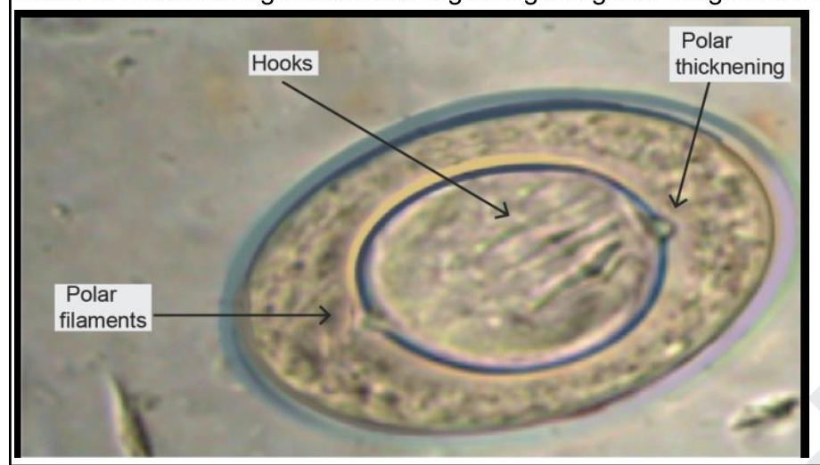
Question 4: Which of the following statements regarding the given image is correct?
- A. Albendazole is the drug of choice
- B. Infection is acquired by ingestion of undercooked freshwater fish
- C. It is the largest tapeworm infecting humans
- D. Majority of infections are asymptomatic in humans (Correct Answer)
Explanation: ***Majority of infections are asymptomatic in humans*** - The image depicts an egg of *Hymenolepis nana*, also known as the **dwarf tapeworm**, identifiable by its characteristic **polar filaments** and **polar thickenings** on the inner membrane. - While heavy infections can cause symptoms, most *Hymenolepis nana* infections are **asymptomatic** or present with only mild, nonspecific gastrointestinal complaints. *Infection is acquired by ingestion of undercooked freshwater fish* - Infection with *Hymenolepis nana* is typically acquired through the **ingestion of eggs** directly from contaminated food or water, or via **fecally-contaminated hands**. - Ingestion of undercooked freshwater fish is associated with trematode (fluke) infections like **Clonorchis sinensis** or **Opisthorchis viverrini**, not *Hymenolepis nana*. *Albendazole is the drug of choice* - The drug of choice for *Hymenolepis nana* infection is **Praziquantel**, given as a single dose. - While albendazole might be used for some helminth infections, it is **less effective** than praziquantel for *Hymenolepis nana*. *It is the largest tapeworm infecting humans* - *Hymenolepis nana* is known as the **dwarf tapeworm** and is generally the **smallest tapeworm** that infects humans, typically measuring a few centimeters in length. - The largest tapeworm infecting humans is *Diphyllobothrium latum* (fish tapeworm), which can reach lengths of several meters.
Question 5: Which of the following are acid-fast staining organisms? 1. Nocardia 2. Mycobacterium leprae 3. Actinomyces 4. Cryptosporidium parvum 5. Isospora belli
- A. 1,2,3
- B. 1,2,3,4,5
- C. 1,2,4,5 (Correct Answer)
- D. 3,4,5
Explanation: ***1,2,4,5*** - **Nocardia**, **Mycobacterium leprae**, **Cryptosporidium parvum**, and **Isospora belli** all exhibit acid-fast properties, meaning they retain carbolfuchsin stain even after decolorization with acid alcohol due to the presence of mycolic acid in their cell walls or unique cyst structures. - This characteristic is crucial for their identification in clinical microbiology and distinguishes them from many other microorganisms. *1,2,3* - This option incorrectly includes **Actinomyces** as an acid-fast organism. **Actinomyces** are Gram-positive, filamentous bacteria that are **not acid-fast**. - While Nocardia and Mycobacterium leprae are acid-fast, the inclusion of Actinomyces makes this choice incorrect. *1,2,3,4,5* - This option is incorrect because it includes **Actinomyces** as an acid-fast organism, which is not true. - **Actinomyces** are Gram-positive, non-acid-fast bacteria, differentiating them from the other listed organisms that do possess acid-fast properties. *3,4,5* - This option is incorrect because it excludes **Nocardia** and **Mycobacterium leprae**, both of which are prominent acid-fast organisms. - While Cryptosporidium parvum and Isospora belli are acid-fast, the omission of Nocardia and Mycobacterium leprae makes this answer incomplete and incorrect.
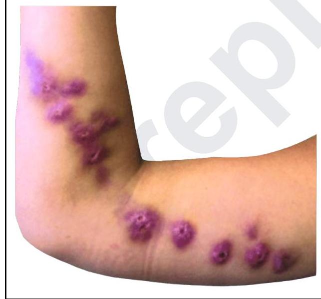
Question 6: A young man presents with skin lesions as shown in the image below. All of the following organisms can spread through dermal and subcutaneous lymphatics, except
- A. Sporothrix schenckii
- B. Staphylococcus aureus (Correct Answer)
- C. Nocardia asteroides
- D. Mycobacterium marinum
Explanation: ***Staphylococcus aureus*** - While *Staphylococcus aureus* can cause various skin infections, it primarily spreads through **direct extension** or the **bloodstream**, not typically through the dermal and subcutaneous lymphatics in a pattern like the one shown. - Infections like cellulitis, abscesses, and impetigo caused by *Staphylococcus aureus* are usually localized or spread via contiguous tissue, rather than forming **linear nodular lesions** along lymphatic channels. *Sporothrix schenckii* - This fungus is a classic cause of **sporotrichosis**, which often presents with **lymphocutaneous spread** following traumatic inoculation. - The image shows **linearly arranged subcutaneous nodules** proximally along the arm, characteristic of lymphatic dissemination, often seen in sporotrichosis. *Nocardia asteroides* - **Nocardia infections** can also cause **lymphocutaneous disease** with a similar appearance to sporotrichosis, especially in immunocompromised individuals. - It can lead to a **chain of subcutaneous nodules and abscesses** tracking along lymphatic vessels from the initial site of infection. *Mycobacterium marinum* - **Mycobacterium marinum** causes **fish tank granuloma** or **swimming pool granuloma** following skin trauma in contaminated water. - It characteristically produces **ascending lymphocutaneous nodules** along lymphatic channels, similar to sporotrichosis, creating a **sporotrichoid pattern**. - The infection typically starts as a papule at the inoculation site and spreads proximally along lymphatics.
Question 7: Which virus can be identified by a PCR method and is endemic to India?
- A. Chikungunya virus (Correct Answer)
- B. Ebola virus
- C. Yellow fever
- D. Hendra virus
Explanation: ***Chikungunya virus*** - The **Chikungunya virus** is a mosquito-borne alphavirus that causes fever, severe joint pain, and rash, and is **endemic to India** and other tropical regions. - Diagnosis is commonly confirmed using **PCR** (polymerase chain reaction) to detect viral RNA in acute samples. *Ebola virus* - The **Ebola virus** causes severe hemorrhagic fever and is primarily prevalent in **Sub-Saharan Africa**, not endemic to India. - While it can be detected by **PCR**, its geographical distribution does not match the endemic criteria for India. *Yellow fever* - **Yellow fever virus** is transmitted by mosquitoes and is endemic to **tropical and subtropical areas of South America and Africa**. - India is not considered an endemic area for yellow fever, though it can be detected by **PCR**. *Hendra virus* - The **Hendra virus** is a zoonotic virus primarily found in **Australia**, transmitted from bats to horses and then to humans. - It is not endemic to India and thus does not fit the criteria of the question.
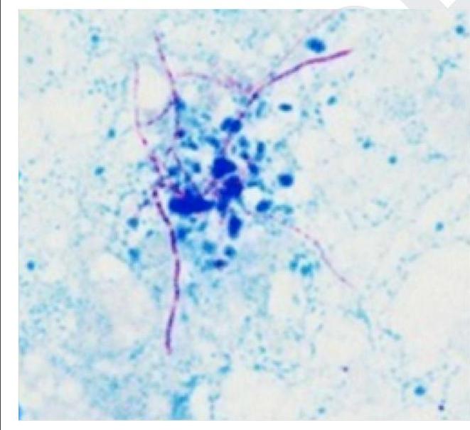
Question 8: Which of the following is a gram-positive organism that shows the following appearance on Ziehl-Neelsen staining?
- A. Nocardia (Correct Answer)
- B. Mycobacterium tuberculosis
- C. Actinomyces
- D. Rhodococcus
Explanation: ***Nocardia*** * The image displays delicate, branching, **filamentous rods** that are stained **red/pink** against a blue background, which is characteristic of partially acid-fast organisms like *Nocardia* on a Ziehl-Neelsen stain. * *Nocardia* species are **gram-positive**, aerobic bacteria that can cause opportunistic infections, particularly in immunocompromised individuals. They are distinguished by their **partial acid-fastness** due to their mycolic acid content, similar to mycobacteria but to a lesser degree. * The characteristic **branching filamentous morphology** combined with partial acid-fastness on Ziehl-Neelsen staining is pathognomonic for *Nocardia*. *Incorrect: Mycobacterium tuberculosis* * While *M. tuberculosis* is **strongly acid-fast** on Ziehl-Neelsen staining (appearing red), it is **not truly gram-positive**—it is gram-variable or weakly gram-positive. * *Mycobacterium* appears as **straight or slightly curved rods**, NOT branching filaments like those shown in the image. *Incorrect: Actinomyces* * *Actinomyces* is a **gram-positive**, filamentous, branching organism that can morphologically resemble *Nocardia*. * However, *Actinomyces* is **NOT acid-fast** and would appear **blue** (not red/pink) on Ziehl-Neelsen staining as it takes up the counterstain. * *Actinomyces* is also anaerobic, whereas *Nocardia* is aerobic. *Incorrect: Rhodococcus* * *Rhodococcus* is a gram-positive organism that can show **partial acid-fastness**, similar to *Nocardia*. * However, *Rhodococcus* typically appears as **coccoid to short rods**, occasionally forming short chains, but does NOT show the extensive **branching filamentous** pattern characteristic of *Nocardia* seen in the image.
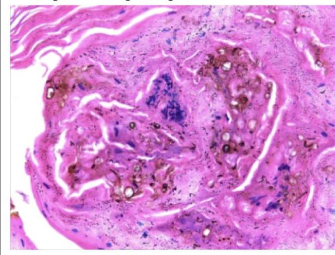
Question 9: A forest worker developed skin lesions over the forearm, which initially started as macules but then became nodules. Histology of the nodule shows the following findings. Which of the following is true regarding this condition?
- A. Angioinvasion is common especially in people with hemolytic anemia
- B. These bodies are formed by engulfment of the dead fungi by the macrophages
- C. It is a dematiaceous fungus (Correct Answer)
- D. Infection commonly spreads to involve tendon, muscle and bone
Explanation: The image displays **chromoblastomycosis**, a fungal infection characterized by **medlar bodies** or **sclerotic bodies**. These are thick-walled, septate, dematiaceous (pigmented) fungal cells that resemble copper pennies. The patient's history of being a forest worker with skin lesions progressing from macules to nodules is consistent with this diagnosis as it's often associated with **traumatic inoculation** from contaminated plant material. ***It is a dematiaceous fungus*** - The image shows **"copper pennies"** or **sclerotic bodies**, which are characteristic of dematiaceous (pigmented) fungi causing chromoblastomycosis. - These fungi contain **melanin** in their cell walls, which contributes to their characteristic dark appearance. - Common causative agents include *Fonsecaea pedrosoi*, *Phialophora verrucosa*, and *Cladophialophora carrionii*. *Angioinvasion is common especially in people with hemolytic anemia* - **Angioinvasion** is not a feature of chromoblastomycosis, which typically remains confined to the **skin and subcutaneous tissue**. - Angioinvasion is characteristic of **mucormycosis** and **aspergillosis**, particularly in immunocompromised patients, not chromoblastomycosis. *These bodies are formed by engulfment of the dead fungi by the macrophages* - The **sclerotic bodies** are **living fungal cells** in their tissue-specific form, not dead fungi engulfed by macrophages. - They are a distinct morphological form of the fungus, adapting to growth within the host tissue, and are **actively pathogenic**. - These thick-walled structures allow the fungus to persist in tissue and resist host defenses. *Infection commonly spreads to involve tendon, muscle and bone* - Chromoblastomycosis causes **chronic, localized infections** primarily of the **skin and subcutaneous tissue**. - While local tissue destruction can occur, **deep invasion** into tendons, muscles, or bones is **rare** and occurs only in severe, long-standing cases. - The infection typically remains confined to cutaneous and subcutaneous layers without dissemination.
Question 10: The viruses of the Filoviridae family like Ebola and Marburg resemble which of the following morphologies?
- A. Brick shaped
- B. Bullet shaped
- C. Spherical
- D. Filamentous (Correct Answer)
Explanation: ***Filamentous*** - Viruses in the **Filoviridae family**, including **Ebola** and **Marburg**, are characterized by their distinct **long, filamentous shape**. - This morphology is a key distinguishing feature visible under electron microscopy, contributing to their namesake ("filo" meaning "thread-like"). *Brick shaped* - **Brick-shaped morphology** is characteristic of **Poxviridae**, such as the **variola virus** (smallpox). - This shape is distinctly different from the thread-like structure of filoviruses. *Bullet shaped* - **Bullet-shaped viruses** are typical of the **Rhabdoviridae family**, which includes the **rabies virus**. - This shape is consistent and easily recognizable for this family, contrasting with the much longer and flexible filaments of filoviruses. *Spherical* - **Spherical morphology** is common among many virus families, including **influenza virus** (Orthomyxoviridae) and **human immunodeficiency virus (HIV)** (Retroviridae). - While many viruses are roughly spherical, filoviruses are specifically known for their elongated, non-spherical appearance.