All SubjectsAnatomy (15)Anesthesiology (1)Biochemistry (8)Community Medicine (11)Dermatology (7)ENT (2)Forensic Medicine (7)Internal Medicine (16)Microbiology (13)Obstetrics and Gynecology (9)Ophthalmology (3)Orthopaedics (2)Pathology (16)Pediatrics (8)Pharmacology (14)Physiology (11)Psychiatry (4)Radiology (5)Surgery (12)
Q11
A 50-year-old male patient presented with left -sided hemiparesis. Damage to which part of the internal capsule leads to this presentation?
Q12
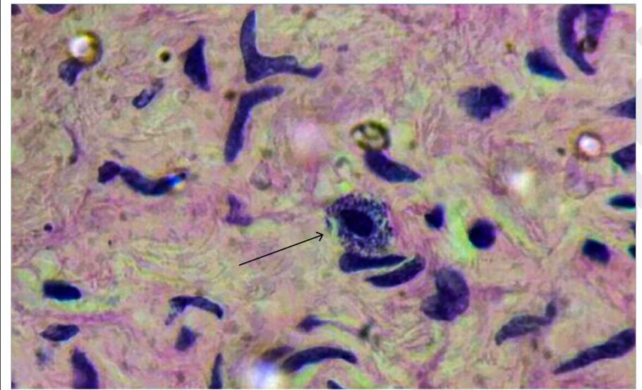
Identify the cell marked in the image below
Q13
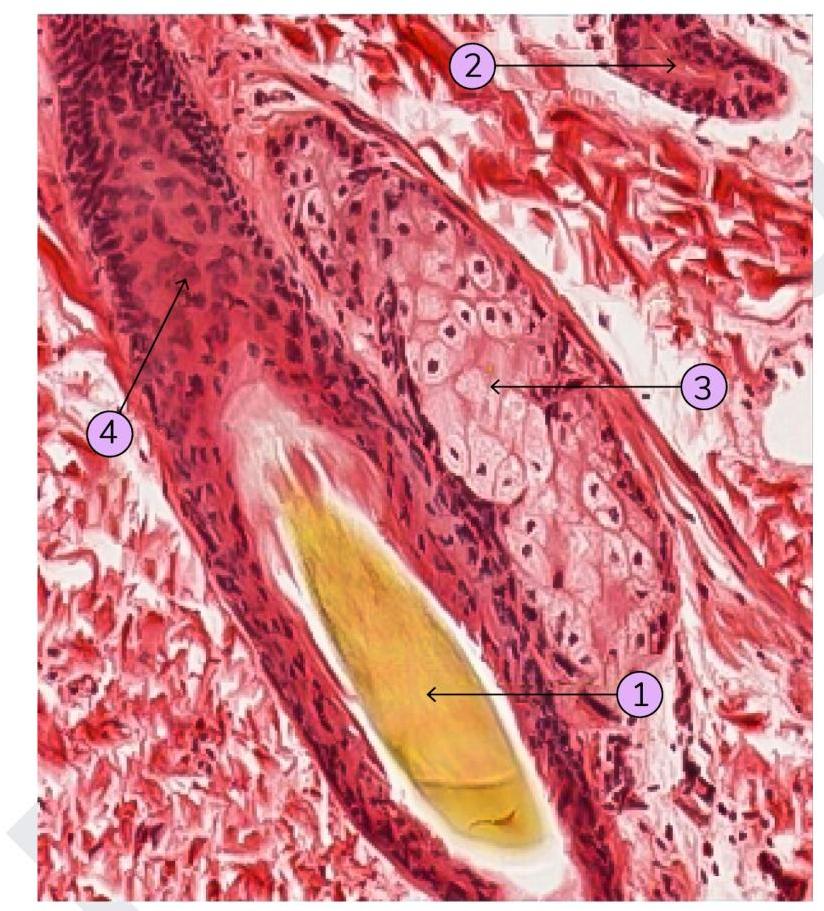
Identify which of the following structure is a sebaceous gland:
Q14
Fingerprint first develops in how many weeks of intrauterine life?
Q15
Froment's sign (book test) is used to assess the function of adductor pollicis. Which of the following nerves supplies this muscle?