INI-CET 2017 — Radiology
5 Previous Year Questions with Answers & Explanations
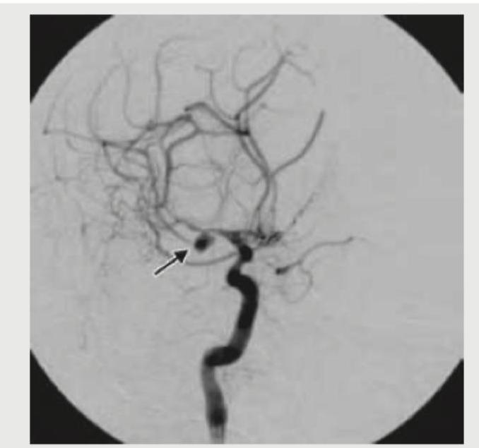
The digital subtraction angiography given below shows? (AIIMS Nov 2017)
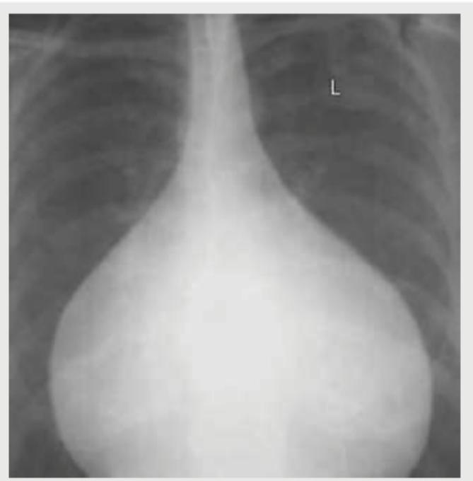
Which is not a finding of the Chest X-ray shown below? (AIIMS May 2017)
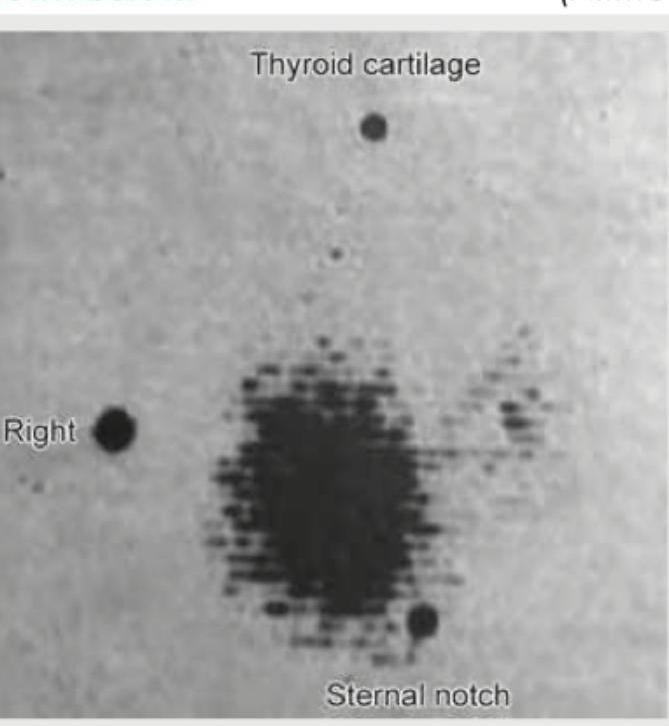
Comment on the diagnosis from the thyroid scan shown below. (AIIMS May 2017)
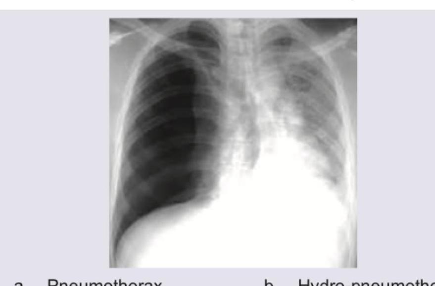
A patient presented with sudden onset difficulty in breathing with RR 28/min, normal blood pressure. X-ray was taken which is given below. What is the diagnosis?
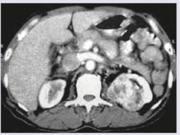
A 50-year-old man went for his annual medical check-up. CT scan is shown below. Diagnosis is?
INI-CET 2017 - Radiology INI-CET Practice Questions and MCQs
Question 1: The digital subtraction angiography given below shows? (AIIMS Nov 2017)
- A. Intra-cranial pseudo-aneurysm
- B. Carotid-cavernous fistula (Correct Answer)
- C. Angiofibroma tumor blush
- D. Vein of Galen malformation
Explanation: ***Carotid-cavernous fistula*** - The image shows early opacification of the **cavernous sinus** and cerebral veins (indicated by the arrow) directly from the internal carotid artery, which is characteristic of a carotid-cavernous fistula. - This high-flow shunt between the arterial and venous systems leads to venous congestion and often presents with **pulsatile exophthalmos**, chemosis, and ophthalmoplegia. *Intra-cranial pseudo-aneurysm* - A pseudo-aneurysm typically appears as a sac-like outpouching from an artery, lacking a true vessel wall, and would not show direct early venous filling. - Pseudo-aneurysms are often caused by **trauma** or infection and may present with signs of hemorrhage if ruptured. *Angiofibroma tumor blush* - An angiofibroma would appear as a **hypervascular mass** with a dense, persistent tumor blush on angiography, but it would not show early direct shunting into veins. - These tumors are typically found in the nasopharynx and present with symptoms like **epistaxis** and nasal obstruction. *Vein of Galen malformation* - A **Vein of Galen malformation** is a developmental anomaly involving an arteriovenous fistula that drains into a dilated median prosencephalic vein, often presenting in infants with high-output heart failure. - While it is an arteriovenous malformation, its typical location and presentation are different from the findings shown in this image, which demonstrates shunting in the region of the cavernous sinus.
Question 2: Which is not a finding of the Chest X-ray shown below? (AIIMS May 2017)
- A. Narrow vascular pedicle (Correct Answer)
- B. Pulmonary venous hypertension
- C. Increased CT ratio
- D. Acute cardiophrenic angle
Explanation: ***Narrow vascular pedicle*** - The image shows a **markedly enlarged cardiac silhouette** with a **"flask-shaped" or "water bottle" heart**, classic for pericardial effusion. In this condition, the vascular pedicle (the mediastinal structures above the heart including the aorta and superior vena cava) is typically **normal to widened** due to venous congestion. - A **narrow vascular pedicle** is characteristically seen in hypovolemia, dehydration, or certain congenital heart diseases with reduced pulmonary blood flow (e.g., tetralogy of Fallot). This finding is **NOT present** in this radiograph. - This is the **most definitively absent finding** among the options listed. *Pulmonary venous hypertension* - The chest X-ray shows prominent pulmonary vascular markings, particularly in the upper lobes, indicative of **cephalization of vessels**, a classic sign of pulmonary venous hypertension. - This occurs due to increased pressure in the pulmonary veins, commonly seen in congestive heart failure or significant pericardial effusion with cardiac tamponade physiology. - This finding **IS present** on the radiograph. *Increased CT ratio* - The **cardiothoracic (CT) ratio** is markedly increased, with the cardiac silhouette clearly exceeding 50% of the thoracic diameter. This indicates **cardiomegaly**, which can result from cardiac chamber enlargement or pericardial effusion. - The extreme enlargement seen here, with the globular "water bottle" configuration, is pathognomonic for large pericardial effusion. - This finding **IS present** on the radiograph. *Acute cardiophrenic angle* - The cardiophrenic angles (the angles formed where the heart border meets the diaphragm laterally) appear **blunted or obtuse** rather than acute (sharp). - While the term "acute cardiophrenic angle" typically refers to the normal sharp angle seen in healthy individuals, the phrasing here is ambiguous. The **angles themselves are present but blunted**, not acute. - However, compared to "narrow vascular pedicle," the blunting of these angles IS a radiographic finding that can be observed, even if abnormal. The vascular pedicle narrowness is completely absent.
Question 3: Comment on the diagnosis from the thyroid scan shown below. (AIIMS May 2017)
- A. Papillary cancer thyroid
- B. Hypersecreting adenoma (Correct Answer)
- C. Grave's disease
- D. Lateral aberrant thyroid
Explanation: ***Hypersecreting adenoma*** - The thyroid scan shows a **single, intensely "hot" nodule**, indicating increased uptake of the radioactive tracer in a localized area. - This pattern is characteristic of a **hyperfunctioning thyroid adenoma**, where the adenoma produces hormones independently, leading to suppression of the rest of the normal thyroid tissue (which appears "cold" or has reduced uptake). *Papillary cancer thyroid* - Papillary thyroid cancer typically appears as a **"cold" nodule** on a thyroid scan, meaning it has reduced or no tracer uptake. - Malignant nodules generally do **not accumulate iodine** as efficiently as normal thyroid tissue or hyperfunctioning benign nodules. *Grave's disease* - Grave's disease presents with **diffuse uptake of the tracer** throughout the entire thyroid gland, not a single localized hot nodule. - The entire gland is generally enlarged and hyperactive, showing **symmetrically increased uptake**. *Lateral aberrant thyroid* - A lateral aberrant thyroid refers to **ectopic thyroid tissue** usually found in the neck, often in lymph nodes due to metastatic papillary carcinoma. - While it involves thyroid tissue outside the normal gland, it wouldn't typically manifest as a single hyperfunctioning nodule within the main thyroid gland, and most ectopic thyroid tissue would show varying uptake depending on its function or if it's metastatic cancer.
Question 4: A patient presented with sudden onset difficulty in breathing with RR 28/min, normal blood pressure. X-ray was taken which is given below. What is the diagnosis?
- A. Pneumothorax
- B. Hydro-pneumothorax (Correct Answer)
- C. Pleural effusion
- D. Consolidation
Explanation: ***Hydro-pneumothorax*** - The chest X-ray clearly shows a **horizontal air-fluid level** in the right hemithorax, indicating the presence of both air (pneumothorax) and fluid (hydrothorax) within the pleural space. - The patient's sudden onset **difficulty in breathing** and **tachypnea (RR 28/min)** are consistent with significant lung pathology like a hydropneumothorax, which compromises lung function. *Pneumothorax* - A simple pneumothorax would show only **air in the pleural space**, characterized by a visible visceral pleural line and absence of lung markings beyond it. - While there is air present, the prominent **fluid level** rules out a diagnosis of pneumothorax alone. *Pleural effusion* - Pleural effusion presents as a **blunting of the costophrenic angles** and a meniscus sign, where fluid conforms to the shape of the thorax. - This image shows a **straight air-fluid level**, not a typical meniscus, indicating the presence of air in addition to fluid. *Consolidation* - Consolidation refers to the **filling of alveolar spaces with fluid or exudate**, appearing as an opacification within the lung parenchyma. - Consolidations typically do not present with a **horizontal fluid level** in the pleural space; they are intraparenchymal.
Question 5: A 50-year-old man went for his annual medical check-up. CT scan is shown below. Diagnosis is?
- A. Renal cell carcinoma
- B. Renal angiomyolipoma (Correct Answer)
- C. Renal cyst
- D. Rhabdomyosarcoma
Explanation: ***Renal angiomyolipoma*** - The CT scan shows a renal mass with areas of **macroscopic fat density**, which is the hallmark of an angiomyolipoma. - Angiomyolipomas are **benign renal tumors** composed of variable amounts of smooth muscle, vascular tissue, and mature adipose tissue. *Renal cell carcinoma* - While renal cell carcinoma can present as a solid renal mass, it typically does **not contain macroscopic fat**. - It usually enhances heterogeneously with contrast and may show areas of necrosis or hemorrhage, but the presence of fat rules out typical RCC. *Renal cyst* - Renal cysts are typically **simple fluid-filled structures** with very low attenuation values (close to water) and **do not contain solid components or fat**. - They also have thin, imperceptible walls and do not enhance with contrast. *Rhabdomyosarcoma* - Rhabdomyosarcomas are **malignant soft tissue tumors** rarely found in the kidney, and would appear as a solid, often heterogeneous mass on CT. - They do **not contain fat** and are aggressive tumors, often associated with a different patient demographic (e.g., children).