All (115)Anatomy (1)Anatomy (10)Anesthesiology (2)Biochemistry (1)Community Medicine (3)Dermatology (13)ENT (5)Forensic Medicine (3)General Medicine (1)Internal Medicine (7)Internal Medicine (2)Microbiology (10)Obstetrics and Gynecology (3)Ophthalmology (5)Orthopaedics (6)Pathology (9)Pathology (9)Physiology (7)Radiology (5)Surgery (3)Surgery (10)
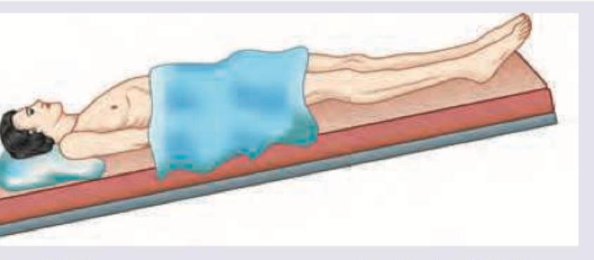
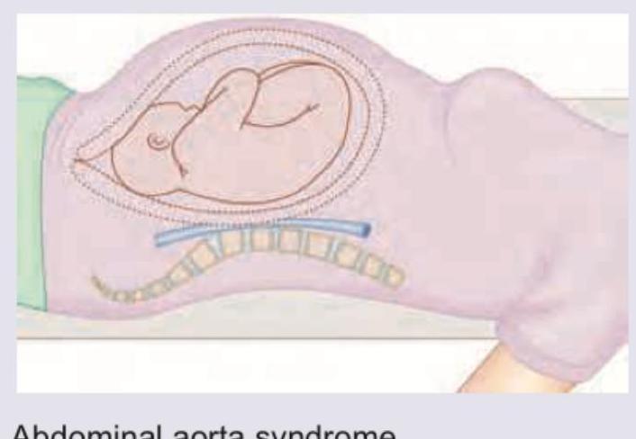
Q61
The position of the patient as shown below is favored for which of the following conditions?