INI-CET 2017 — Microbiology
9 Previous Year Questions with Answers & Explanations
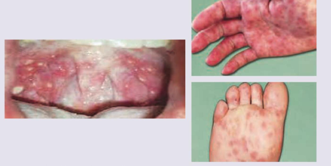
Which causative organism is responsible for this disease?
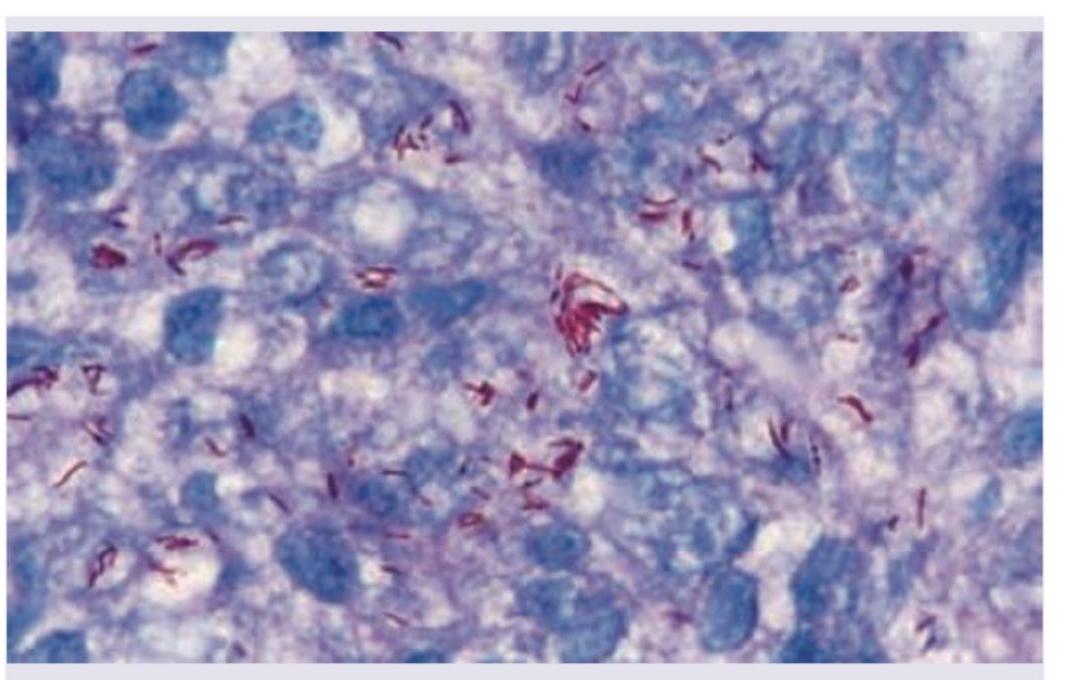
Identify the organism seen in the slide shown below.
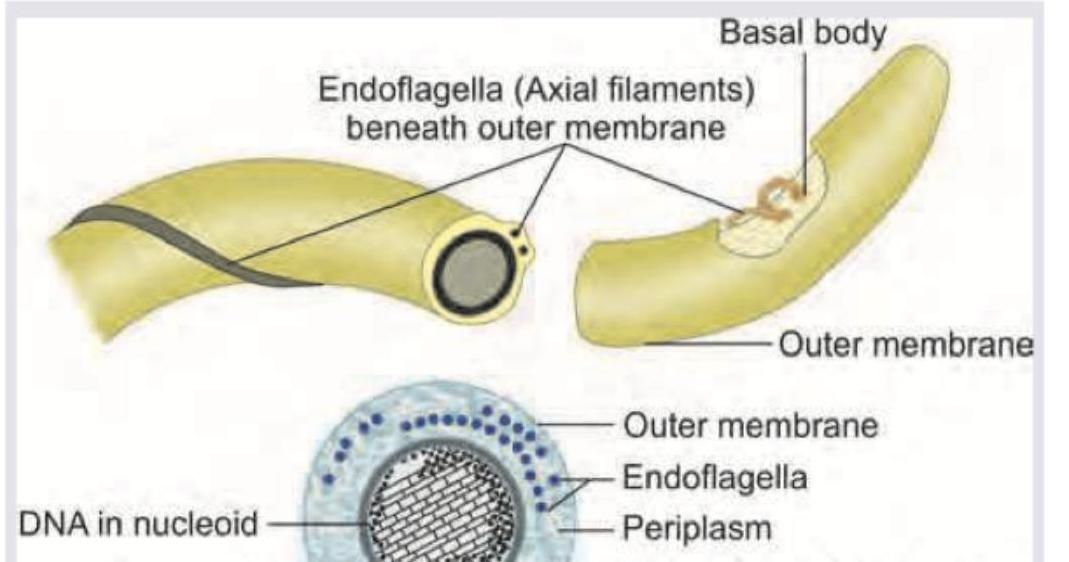
Which of the following bacteria has the flagellar characteristic shown in the image?
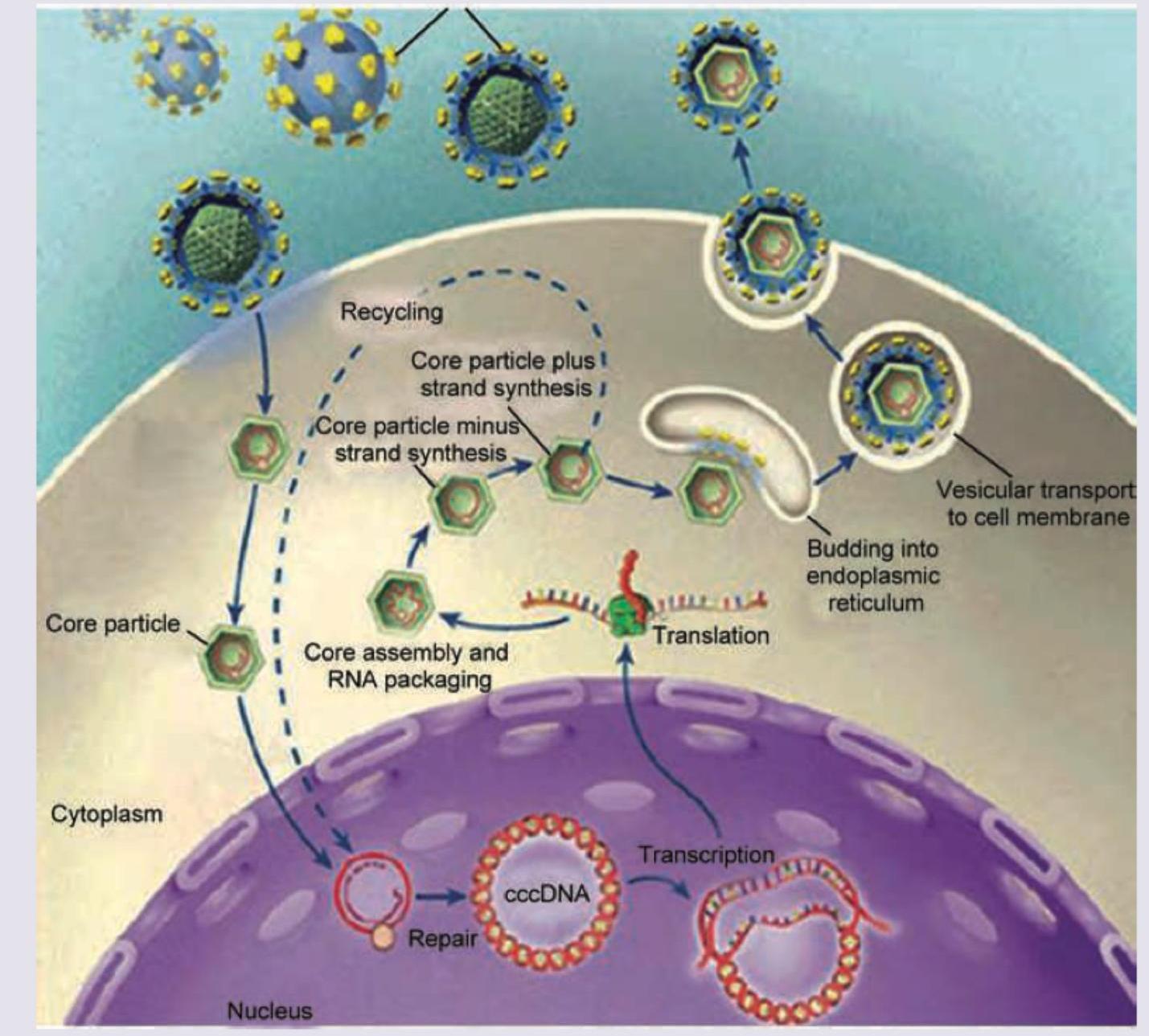
Amongst the four choices, select the virus whose life cycle is shown. (AIIMS Nov 2017)
Identify the fungal organism in this slide stained with Gomori-methenamine silver stain. (AIIMS Nov 2017)
Which of the following parasite's life cycle is shown below?
Which of the following fits into the life cycle of the picture given below? (AIIMS May 2017)
The image shown below shows egg of? (AIIMS May 2017)
Identify the organism shown below. (AIIMS May 2017)
INI-CET 2017 - Microbiology INI-CET Practice Questions and MCQs
Question 1: Which causative organism is responsible for this disease?
- A. Coxsackie virus (Correct Answer)
- B. Human herpes virus 7
- C. Pox virus
- D. Molluscum contagiosum virus
Explanation: ***Coxsackie virus*** - The images show typical lesions of **Hand-Foot-and-Mouth Disease (HFMD)**, characterized by **oral ulcers** (herpangina) and a vesiculopapular rash on the **palms, soles**, and sometimes buttocks. - HFMD is most commonly caused by **Coxsackie virus A16** and other enteroviruses. *Human herpes virus 7* - This virus is primarily associated with **roseola infantum (exanthem subitum)**, characterized by rapid onset of high fever followed by a rash after the fever breaks. - The rash is typically maculopapular and found on the trunk, not primarily on the palms, soles, and mouth as seen in the image. *Pox virus* - Poxviruses cause diseases like **smallpox** and **molluscum contagiosum**, which present with different types of lesions. - **Smallpox** lesions are deep-seated, painful pustules that evolve synchronously, and **molluscum contagiosum** manifests as pearly, umbilicated papules, neither matching the depicted oral and acral rash. *Molluscum contagiosum virus* - Molluscum contagiosum virus (MCV) is a type of **poxvirus** that causes the skin infection **molluscum contagiosum**. - Its characteristic lesions are **dome-shaped, pearly papules with central umbilication**, which are not consistent with the vesicular lesions and oral ulcers shown in the image.
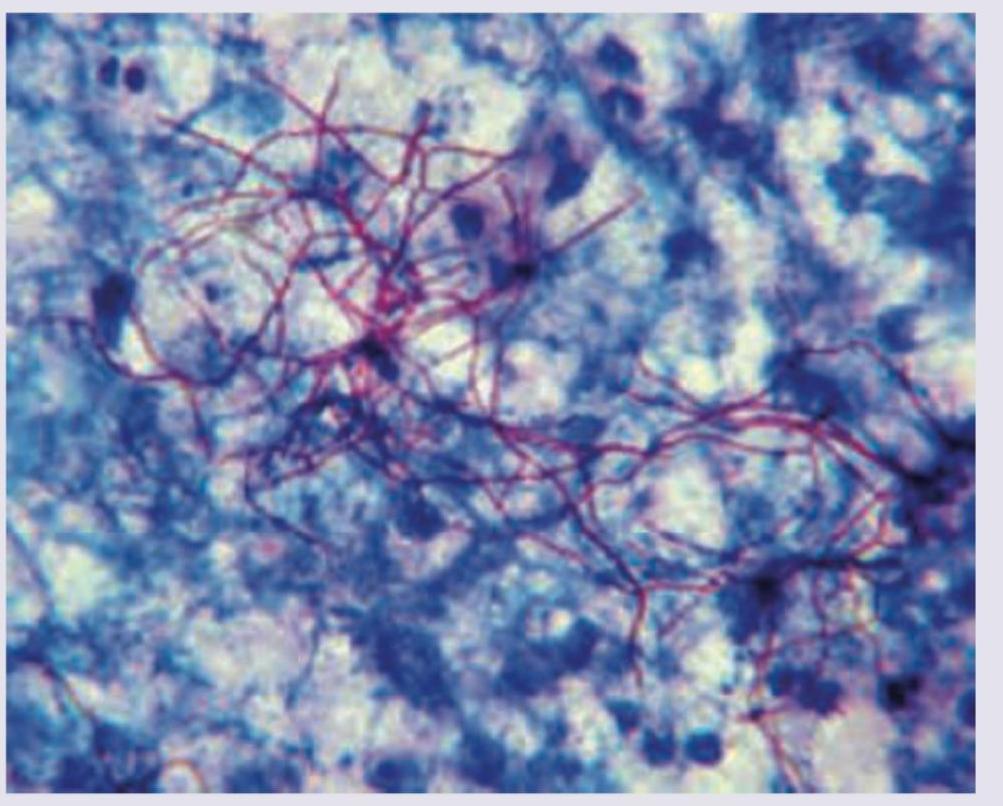
Question 2: Identify the organism seen in the slide shown below.
- A. Streptococcus pyogenes
- B. M. tuberculosis
- C. Nocardia asteroides (Correct Answer)
- D. Corynebacterium diphtheriae
Explanation: ***Nocardia asteroides*** - The image exhibits **branching, beaded, filamentous rods** that stain **acid-fast**, key characteristics of *Nocardia*. - These morphological features, especially the irregular acid-fast staining and branching, differentiate it from other bacteria. *Streptococcus pyogenes* - *Streptococcus pyogenes* are Gram-positive cocci typically arranged in **chains**, showing none of the filamentous or branching forms seen in the image. - They are **not acid-fast** and would not appear as pink/red rods on an acid-fast stain. *M. tuberculosis* - While *M. tuberculosis* is **acid-fast** and appears as red rods, it typically presents as **straight or slightly curved rods**, not the extensively branching, beaded filaments observed. - It does not form the complex filamentous structures characteristic of *Nocardia*. *Corynebacterium diphtheriae* - *Corynebacterium diphtheriae* are Gram-positive, **club-shaped rods** that often assume V or L forms (palisades), and are not acid-fast. - This bacterium does not exhibit the extensive branching or beaded appearance characteristic of the organism in the image.
Question 3: Which of the following bacteria has the flagellar characteristic shown in the image?
- A. Helicobacter pylori
- B. Mycobacterium tuberculosis
- C. Leptospira icterohaemorrhagicae (Correct Answer)
- D. Vibrio cholerae
Explanation: ***Leptospira icterohaemorrhagicae*** - The image displays **endoflagella** (axial filaments) located beneath the **outer membrane**, which is characteristic of **spirochetes**. - *Leptospira icterohaemorrhagicae* is a spirochete and possesses these internal flagella, allowing for its distinctive corkscrew motility. *Helicobacter pylori* - *Helicobacter pylori* is a gram-negative bacterium that typically has **lophotrichous flagella** (multiple flagella at one pole), which are external, not internal. - Its motility is crucial for penetrating the gastric mucus, but it does not utilize endoflagella. *Mycobacterium tuberculosis* - *Mycobacterium tuberculosis* is a non-motile bacterium and **lacks flagella** altogether. - Its cell wall structure, rich in mycolic acid, contributes to its pathogenicity and resistance but not to motility via flagella. *Vibrio cholerae* - *Vibrio cholerae* is a gram-negative bacterium that possesses a **single polar flagellum** (monotrichous), which is external. - This external flagellum is essential for its motility in aquatic environments and in the human intestine.
Question 4: Amongst the four choices, select the virus whose life cycle is shown. (AIIMS Nov 2017)
- A. Hepatitis B (Correct Answer)
- B. Herpes simplex
- C. HIV
- D. Influenza
Explanation: ***Hepatitis B*** - The image clearly depicts the formation of **cccDNA (covalently closed circular DNA)** in the host cell nucleus, a unique and defining characteristic of the Hepatitis B virus life cycle. - The process of **reverse transcription** (synthesis of DNA from an RNA template) occurring within the core particle in the cytoplasm, followed by its movement to the nucleus to form cccDNA, is specific to Hepatitis B. *Herpes simplex* - Herpes simplex viruses are **DNA viruses** that replicate entirely within the nucleus, and their replication does not involve an RNA intermediate or the formation of cccDNA. - They also undergo **budding from the inner nuclear membrane** into the perinuclear space, which differs from the ER budding shown. *HIV* - HIV is a **retrovirus** that utilizes reverse transcriptase to convert its RNA genome into DNA, which then integrates into the host genome. However, it does not form cccDNA. - While it does involve reverse transcription, the overall replication strategy, particularly the integration step and the budding from the cell membrane, differs significantly from the depicted process. *Influenza* - Influenza is an **RNA virus** that replicates in the nucleus (unlike most RNA viruses) but does not involve a DNA intermediate or cccDNA formation. - Its replication cycle involves transcription of viral RNA into mRNA and replication of viral RNA genomes, which is distinct from the complex DNA to RNA to DNA cycle shown.
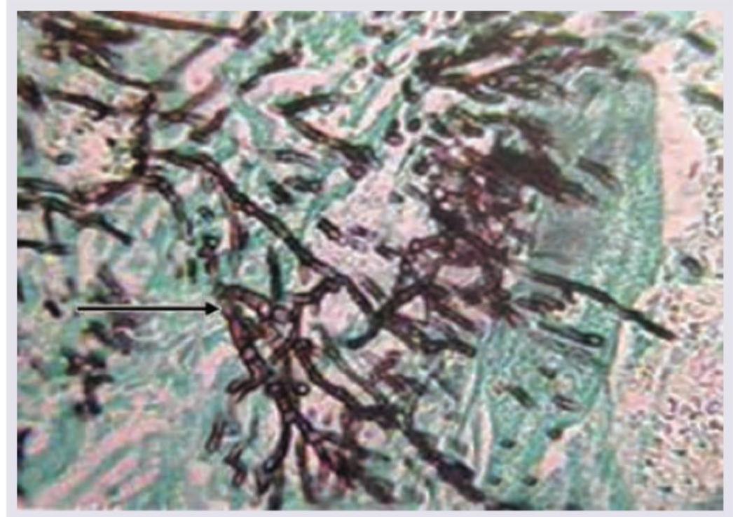
Question 5: Identify the fungal organism in this slide stained with Gomori-methenamine silver stain. (AIIMS Nov 2017)
- A. Acute angle branching, rhizopus
- B. Right angle branching, aspergillus
- C. Acute angle branching, aspergillus (Correct Answer)
- D. Right angle branching, mucor
Explanation: **Acute angle branching, aspergillus** - The image shows **septate hyphae** with **acute angle (45-degree) branching**, which is characteristic of *Aspergillus* species. - While *Aspergillus* can be identified by conidial heads in culture, in tissue sections stained with Gomori-methenamine silver (GMS) stain, these morphological features are key. *Acute angle branching, rhizopus* - **Rhizopus** species typically exhibit **non-septate or sparsely septate hyphae** with **irregular branching**, often at wide angles. - This morphology differs from the regularly septate, acutely branching hyphae seen in the image. *Right angle branching, aspergillus* - **Aspergillus** hyphae characteristically show **acute angle (45-degree) branching**, not right-angle branching. - The hyphae are also **septate**, a feature that is clearly visible in the image. *Right angle branching, mucor* - **Mucor** species, like other Mucorales (e.g., *Rhizopus*, *Lichtheimia*), are known for their **non-septate or sparsely septate hyphae** with **wide-angle (often right-angle) branching**. - The hyphae in the image are clearly septate and branch acutely, ruling out *Mucor*.
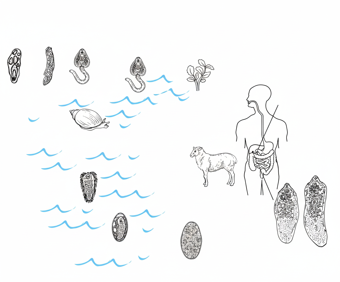
Question 6: Which of the following parasite's life cycle is shown below?
- A. Fasciola hepatica (Correct Answer)
- B. Clonorchis sinensis
- C. Paragonimus westermani
- D. Schistosoma japonicum
Explanation: ***Fasciola hepatica (Liver Fluke)*** - The diagram clearly illustrates the key stages of the Fasciola hepatica life cycle, including **encystation in the duodenum**, migration to the **liver**, and the presence of adult worms in the **bile duct** of humans and animals - The presence of **sheep and cattle** as definitive hosts, and the involvement of **freshwater snails** (Lymnaea species) as intermediate hosts, are characteristic features of *Fasciola hepatica* - **Eggs escaping in feces** into water where miracidia develop and infect snails, followed by cercaria forming **metacercaria on aquatic vegetation** (eaten by herbivores), is pathognomonic of this fluke *Clonorchis sinensis* - This is the **Chinese liver fluke** that also inhabits bile ducts, but uses **freshwater fish** (not vegetation) as the second intermediate host - Humans acquire infection by eating **raw or undercooked fish**, not vegetation *Paragonimus westermani* - This is the **lung fluke** found in the lungs (not liver/bile ducts) - Uses **freshwater crabs or crayfish** as second intermediate hosts - Life cycle does not involve grazing animals or vegetation *Schistosoma japonicum* - This is a **blood fluke** (not a liver fluke) causing schistosomiasis - Cercaria penetrate through **intact skin** (not ingested) - Adult worms live in **mesenteric veins**, not bile ducts - No metacercarial encysted stage on vegetation
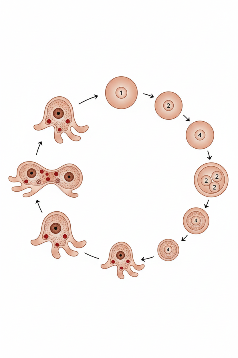
Question 7: Which of the following fits into the life cycle of the picture given below? (AIIMS May 2017)
- A. Entamoeba histolytica (Correct Answer)
- B. Giardia lamblia
- C. Balantidium coli
- D. Entamoeba coli
Explanation: ***Entamoeba histolytica*** - The diagram illustrates the characteristic life cycle of *Entamoeba histolytica*, showing the **trophozoite** stage with pseudopodia, **binary fission**, **encystment** forming a protective cyst wall, and **excystation** producing metacystic amoebae. - Key identifying features include the **4-nucleate mature cyst** and the presence of **chromatoid bars** with rounded ends, which are pathognomonic for this parasite's life cycle. *Giardia lamblia* - This is a **flagellated protozoan** with a distinctive **pear-shaped trophozoite** containing two nuclei and eight flagella, completely different from the amoeboid forms shown. - The life cycle involves **binucleate cysts** and does not include pseudopodial movement or the amoeboid stages depicted in the diagram. *Balantidium coli* - This is a **ciliated protozoan** (the largest intestinal parasite) with a characteristic **kidney-shaped macronucleus** and numerous cilia for motility. - The trophozoite is **oval-shaped** with cilia covering the surface, lacking the pseudopodial extensions and amoeboid characteristics shown in the life cycle. *Entamoeba coli* - While also an amoeba, *E. coli* forms **8-nucleate mature cysts** (compared to 4-nucleate in *E. histolytica*) and has **chromatoid bars with splinter-like pointed ends**. - This non-pathogenic amoeba has a similar but distinct life cycle with different cyst morphology and nuclear characteristics than those depicted.
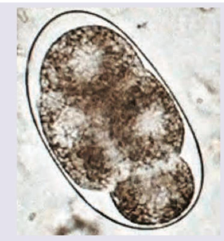
Question 8: The image shown below shows egg of? (AIIMS May 2017)
- A. A. duodenale
- B. E. vermicularis
- C. A. lumbricoides (Correct Answer)
- D. Strongyloides stercoralis
Explanation: ***A. lumbricoides*** - The image displays a **mammillated outer layer** and an **oval shape**, characteristic features of a fertilized *Ascaris lumbricoides* egg. - The internal content shows a **developing embryo** (morula stage), which is typical for newly passed *Ascaris* eggs. *A. duodenale* - The eggs of *Ancylostoma duodenale* (hookworm) are typically **oval or ellipsoidal**, have a **thin, transparent shell**, and contain a **segmented ovum** (usually 2-8 cells) when passed in stool. - They lack the distinctive **thick, mammillated outer layer** seen in the image. *E. vermicularis* - *Enterobius vermicularis* (pinworm) eggs are **D-shaped** or **asymmetrically ovoid**, with one side flattened. - They possess a **thin, smooth shell** and contain a **larva**, which distinguishes them from the egg shown. *Strongyloides stercoralis* - *Strongyloides stercoralis* eggs are rarely seen in stool samples because they usually **hatch within the intestine**, releasing **rhabditiform larvae**. - When present, they are **oval, thin-shelled**, and contain a **partially developed larva**.
Question 9: Identify the organism shown below. (AIIMS May 2017)
- A. Nocardia (Correct Answer)
- B. Mycobacterium tuberculosis
- C. Mycobacterium leprae
- D. Actinomyces
Explanation: ***Nocardia*** - The image displays **branching, filamentous, gram-positive rods** that exhibit a **beaded appearance**, which is characteristic of *Nocardia* species. - *Nocardia* are **partially acid-fast** and can cause opportunistic infections, particularly in immunocompromised individuals. *Mycobacterium tuberculosis* - *Mycobacterium tuberculosis* appears as **rod-shaped bacilli** that are **strongly acid-fast** due to their high mycolic acid content, but they do not typically form the long, branching filaments seen in the image. - While they can form cords, these are not the extensive mycelial-like structures shown. *Mycobacterium leprae* - *Mycobacterium leprae* are **acid-fast bacilli** that typically appear in compact bundles ("globi") within host cells, not as branching filamentous structures. - This organism primarily causes leprosy and is difficult to culture in vitro. *Actinomyces* - *Actinomyces* species are also **branching, filamentous gram-positive bacteria**, but they are **not acid-fast**. - While they form characteristic "sulfur granules" in tissue, the image is a stain showing individual organisms, and the acid-fast appearance rules out *Actinomyces*.