INI-CET 2017 — ENT
3 Previous Year Questions with Answers & Explanations
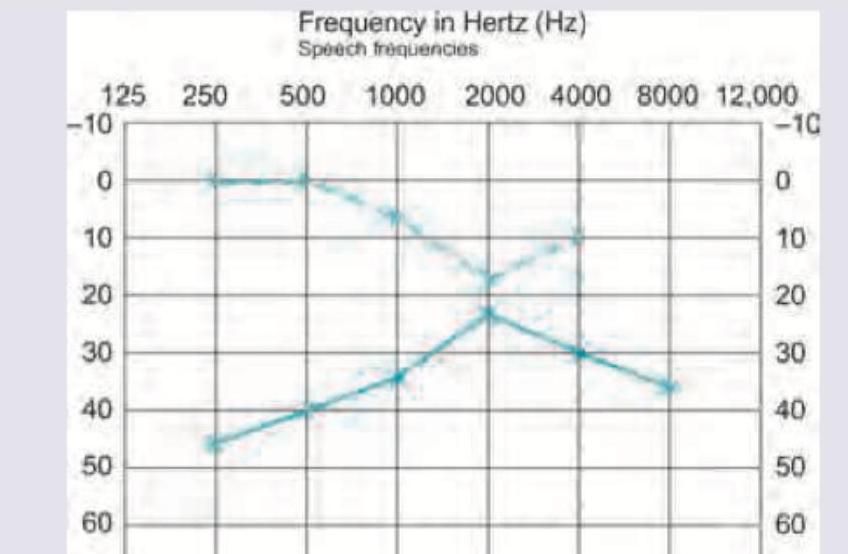
Which of the following is the diagnosis of the audiogram shown below?
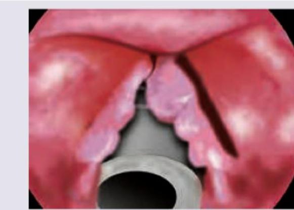
The following image shows:
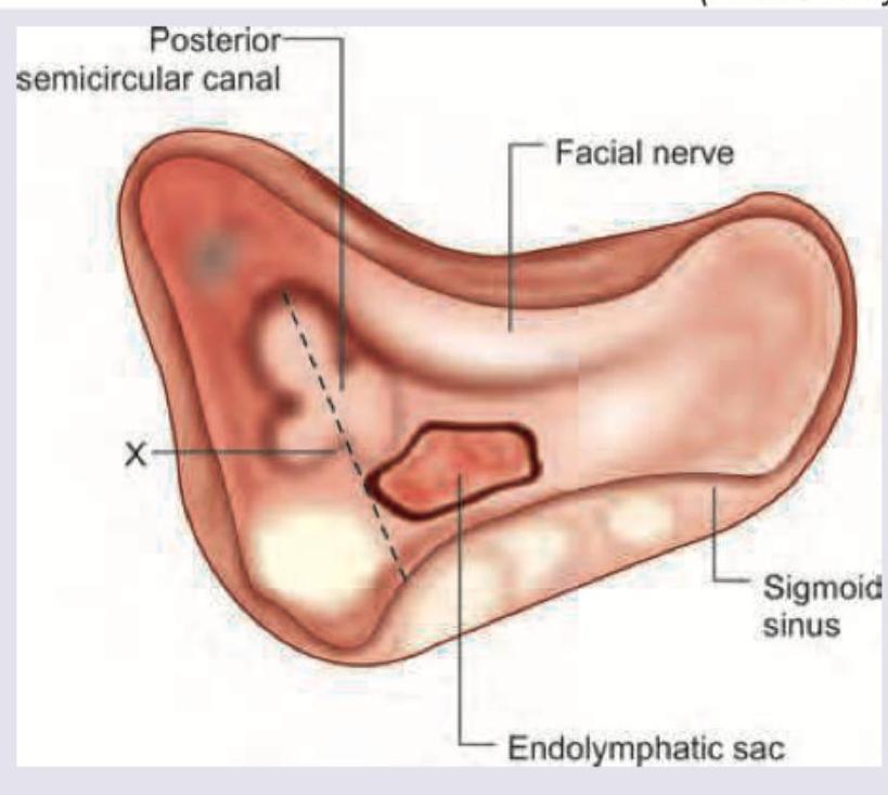
Identify the line shown in the given image:
INI-CET 2017 - ENT INI-CET Practice Questions and MCQs
Question 1: Which of the following is the diagnosis of the audiogram shown below?
- A. Otosclerosis
- B. Ototoxicity
- C. Meniere's disease
- D. Noise induced hearing loss (Correct Answer)
Explanation: ***Noise induced hearing loss*** - The audiogram shows a classic **'noise notch'**, characterized by a dip in hearing at **3000-6000 Hz**, with recovery at 8000 Hz, reflecting damage to hair cells in the cochlea from excessive noise exposure. - Both air conduction (solid line) and bone conduction (dashed line) thresholds are depressed in the same pattern, indicating a **sensorineural hearing loss**. *Otosclerosis* - Otosclerosis typically causes a **conductive hearing loss**, meaning air conduction thresholds would be significantly worse than bone conduction thresholds, showing an **air-bone gap**. - It often results in a characteristic **Carhart notch** (a dip at 2000 Hz) in bone conduction, but this audiogram shows sensorineural loss without a significant air-bone gap. *Ototoxicity* - Ototoxicity usually results in a **high-frequency sensorineural hearing loss**, often affecting frequencies above 4000 Hz first, and typically shows a more gradual, sloping loss rather than a sharp notch. - While it is sensorineural, the specific 'notch' pattern seen here is more characteristic of noise exposure. *Meniere's disease* - Meniere's disease classically presents with a **low-frequency sensorineural hearing loss** that can fluctuate, accompanied by **tinnitus, vertigo, and aural fullness**. - The audiogram does not show a low-frequency loss, nor does it typically present with a noise notch.
Question 2: The following image shows:
- A. Respiratory papillomatosis (Correct Answer)
- B. Vocal nodule
- C. Vocal polyp
- D. TB of vocal cords
Explanation: ***Respiratory papillomatosis*** - The image displays multiple **wart-like growths** on the vocal cords, characteristic of **respiratory papillomatosis**, which is caused by the **human papillomavirus (HPV)**. - These lesions often have an **irregular, cauliflower-like appearance** and can recur even after removal, making it a challenging condition to manage. *Vocal nodule* - Vocal nodules are typically **bilateral, symmetrical lesions** located at the junction of the anterior and middle thirds of the vocal cords. - They are usually **smooth, small, and whitish**, resulting from chronic vocal abuse, unlike the irregular and multiple growths seen in the image. *Vocal polyp* - Vocal polyps are typically **unilateral lesions** that can appear as sessile or pedunculated masses on a vocal cord. - They are often **larger than nodules** and may have a reddish or gelatinous appearance, but they usually occur singly, not as multiple diffuse growths like those pictured. *TB of vocal cords* - Tuberculosis of the vocal cords often presents with **ulcerative lesions**, granulomas, or diffuse inflammation, and may be accompanied by other signs of pulmonary TB. - The lesions caused by TB are generally **not papillomatous** or wart-like in nature, differentiating them from the appearance in the image.
Question 3: Identify the line shown in the given image:
- A. Ohngren's line
- B. Weber Ferguson line
- C. Donaldson line (Correct Answer)
- D. Frankfurt line
Explanation: ***Donaldson line*** - The image illustrates the posterior aspect of the petrous temporal bone, showing the relationship between the **facial nerve**, **posterior semicircular canal**, and the **endolymphatic sac**. - The dashed line directly passes superomedial to the **endolymphatic sac** and inferolateral to the **posterior semicircular canal**, which is characteristic of Donaldson's line, used as a surgical landmark. *Ohngren's line* - Ohngren's line is a radiological landmark used in the context of **maxillary sinus tumors**. - It runs from the **medial canthus of the eye** to the **angle of the mandible**, dividing the maxillary sinus. *Weber Ferguson line* - The Weber-Ferguson incision is a **facial incision** used for access to the **maxillary sinuses and midface**. - It is a surgical approach, not an anatomical line delineating internal structures. *Frankfurt line* - The Frankfurt horizontal plane, or Frankfurt line, is an **anthropometric landmark** used in craniometry and orthodontics. - It is defined by the superior border of the **external auditory meatus** and the inferior orbital rim.