INI-CET 2017 — Anatomy
9 Previous Year Questions with Answers & Explanations
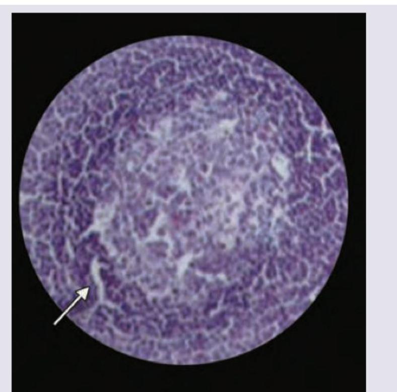
Which of the following area is marked in the histology of lymph node? (AIIMS May 2017)
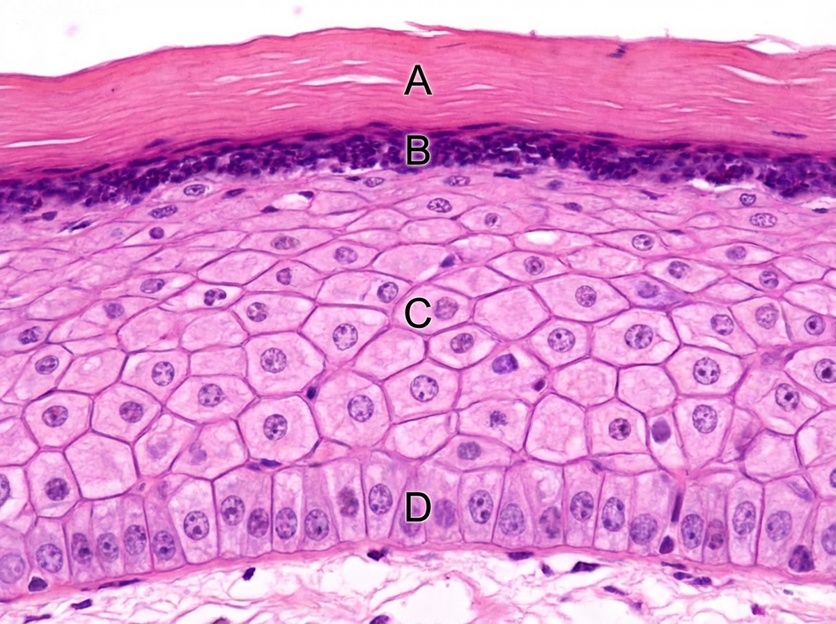
Which of the following layers has abundance of desmosomes? (AIIMS Nov 2017)
An intra-operative photograph of cortical mastoidectomy is shown. Identify the lateral semi-circular canal. (AIIMS Nov 2017)
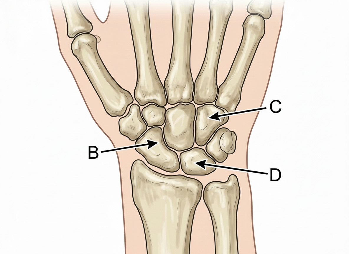
Which of the following bones is the first one to ossify?
Which of the following structures will help in opening of jaw?
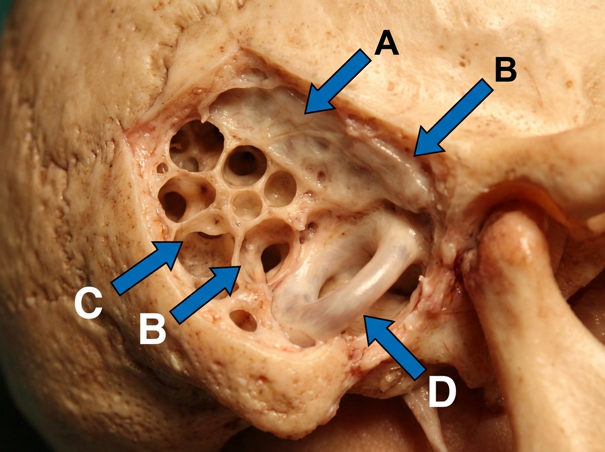
Identify the structure marked by the arrow in this skull base image:
Which of the following has holocrine secretion?
Which of the following proteins are not seen in the region marked in the image?
Which of the following is the constituent of the marked area in the given electron microscope picture of the muscle?
INI-CET 2017 - Anatomy INI-CET Practice Questions and MCQs
Question 1: Which of the following area is marked in the histology of lymph node? (AIIMS May 2017)
- A. Mantle zone
- B. Marginal zone
- C. Germinal center (Correct Answer)
- D. Paracortical area
Explanation: ***Germinal center*** - The image illustrates a **germinal center**, characterized by its **lighter staining** and a distinct network of cells (likely follicular dendritic cells) which are responsible for B-cell proliferation and differentiation. - The pointer indicates the surrounding, more basophilic lymphocytes, often seen adjacent to the paler germinal center. *Mantle zone* - The mantle zone surrounds the germinal center and consists of **small, inactive B-lymphocytes** that stain more densely (darker) than the cells within the germinal center. - It would be seen as a darker ring immediately outside the lighter germinal center. *Marginal zone* - The marginal zone is typically found in the **spleen** and is a region of B cells that surrounds the white pulp. - It is not a primary structural component identified within the follicular architecture of a lymph node in the manner depicted. *Paracortical area* - The paracortex is primarily a **T-cell zone**, located between the follicles and the medulla within the lymph node. - It would not exhibit the distinct follicular structure with a light center and surrounding darker cells as shown.
Question 2: Which of the following layers has abundance of desmosomes? (AIIMS Nov 2017)
- A. A
- B. B
- C. C (Correct Answer)
- D. D
Explanation: ***C*** - Layer C represents the **stratum spinosum** (prickle cell layer) of the epidermis, which is characterized by abundant **desmosomes**. - These desmosomes give the cells a **prickly appearance** when stained, as the cells shrink but the desmosomal attachments remain, pulling on the cell membranes. *A* - Layer A represents the **stratum corneum**, which is the outermost layer consisting of flattened, anucleated cells filled with **keratin**. - While desmosomes are present deeper in the epidermis, this layer is primarily involved in **protection** and shedding of dead cells. *B* - Layer B represents the **stratum granulosum**, characterized by cells containing **keratohyalin granules**. - These granules are precursors to keratin, and while desmosomes connect cells here, they are less prominent than in the stratum spinosum. *D* - Layer D represents the **stratum basale** (basal layer), which is the deepest epidermal layer of cuboidal or columnar cells. - These cells are responsible for **cell proliferation** and connect to the basement membrane via **hemidesmosomes**, and to each other via desmosomes, but not as abundantly as in the stratum spinosum.
Question 3: An intra-operative photograph of cortical mastoidectomy is shown. Identify the lateral semi-circular canal. (AIIMS Nov 2017)
- A. A
- B. B
- C. C
- D. D (Correct Answer)
Explanation: ***Correct Option D*** - The diagram shows a simplified view of the temporal bone anatomy post-mastoidectomy, and 'D' clearly points to the position of the **lateral semicircular canal**. - During cortical mastoidectomy, the **lateral semicircular canal** is typically identified in the superior and posterior wall of the mastoid cavity, just above the aditus and facial nerve. - This is a critical landmark during mastoid surgery as injury to it can cause vertigo and hearing loss. *Incorrect Options A, B, and C* - These labels point to other anatomical structures visible in the mastoid cavity during cortical mastoidectomy, such as the tegmen tympani, facial nerve, sigmoid sinus, or other osseous landmarks. - Proper identification of the lateral semicircular canal is essential to avoid iatrogenic injury during surgery.
Question 4: Which of the following bones is the first one to ossify?
- A. A
- B. B
- C. C (Correct Answer)
- D. D
Explanation: ***C*** - The **capitate bone** is the first carpal bone to ossify, typically appearing around **1-3 months** of postnatal life. - As the largest carpal bone, its early ossification serves as an important radiological marker for assessing **skeletal maturity** in pediatric patients. *A* - The **scaphoid bone** ossifies much later, around **5-6 years** of age, making it one of the last carpal bones to develop. - Due to its **retrograde blood supply**, the scaphoid is particularly susceptible to **avascular necrosis** following fractures. *B* - The **trapezoid bone** typically begins ossification between **4-6 years** of age, significantly later than the capitate. - Located between the **trapezium and capitate**, it forms part of the distal carpal row and contributes to **index finger stability**. *D* - The **lunate bone** usually starts ossifying around **2-4 years** of age, making it the second carpal bone to ossify after the capitate. - The lunate is clinically significant as it's commonly affected by **Kienböck's disease** (avascular necrosis) and **perilunate dislocations**.
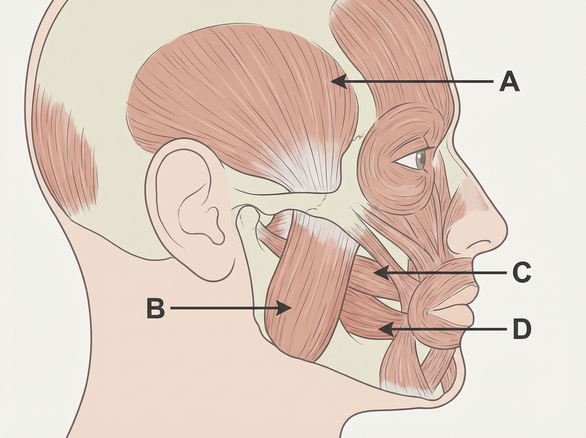
Question 5: Which of the following structures will help in opening of jaw?
- A. A
- B. B
- C. C (Correct Answer)
- D. D
Explanation: ***C*** - Structure C represents the **lateral pterygoid muscle**, which is the primary muscle responsible for **opening the jaw (depression of the mandible)**, as well as protrusion and contralateral excursion. - It is the only muscle of mastication that actively participates in jaw opening. *A* - Structure A appears to be the **medial pterygoid muscle**, which is primarily involved in **elevation of the mandible** (jaw closing) and side-to-side movements. - Its action is antagonistic to jaw opening. *B* - Structure B likely represents the **masseter muscle**, a powerful muscle of mastication that primarily functions to **elevate the mandible** and close the jaw. - It is a strong jaw closer, not an opener. *D* - Structure D points to the **temporalis muscle**, another major muscle of mastication that is responsible for **elevating and retracting the mandible** (closing the jaw). - Its primary actions are in jaw closing, not opening.
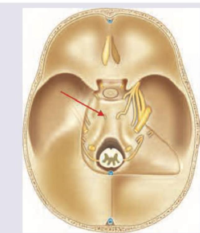
Question 6: Identify the structure marked by the arrow in this skull base image:
- A. Spinal accessory nerve
- B. Vertebral artery (Correct Answer)
- C. Labyrinthine artery
- D. Abducens nerve
Explanation: ***Vertebral artery*** - The arrow indicates the **vertebral artery** as it ascends through the **foramen magnum** into the posterior fossa, appearing as a prominent paired vascular structure lateral to the **medulla oblongata**. - In skull base imaging, vertebral arteries appear as **cylindrical structures** running alongside the brainstem, distinguishable from nerves by their **larger caliber** and **bilateral symmetry** at the craniovertebral junction. *Spinal accessory nerve* - The **spinal accessory nerve (CN XI)** has a much **smaller diameter** than the indicated structure and would appear as a thin nerve bundle coursing toward the **jugular foramen**. - It enters the skull through the foramen magnum but quickly turns laterally toward the **jugular foramen**, not maintaining the vertical course shown by the arrow. *Labyrinthine artery* - The **labyrinthine artery** is a **small branch** of the AICA that enters the **internal acoustic meatus** to supply the inner ear, too small to be clearly visible at this magnification. - It would be located more **anterolaterally** near the cerebellopontine angle, not in the **midline posterior fossa** location indicated by the arrow. *Abducens nerve* - The **abducens nerve (CN VI)** emerges from the **pontomedullary junction** and travels anteriorly through the **cavernous sinus**, located much more **superiorly and anteriorly** than the marked structure. - It would appear as a **thin nerve** rather than the **robust vascular structure** indicated, and would not be visible at the **foramen magnum level**.
Question 7: Which of the following has holocrine secretion?
- A. Sebaceous gland (Correct Answer)
- B. Salivary gland
- C. Mammary gland
- D. Sweat gland
Explanation: ***Sebaceous gland*** - Sebaceous glands use **holocrine secretion**, in which the **entire cell disintegrates** to release its secretory product (sebum) - Cells progressively accumulate lipid in the cytoplasm, undergo degeneration, and ultimately lyse completely to form the secretion - This is the **only example of holocrine secretion** in the human body - Histologically, sebaceous glands show large, pale, lipid-filled cells arranged in lobules with a central duct opening into the hair follicle *Sweat gland* - Eccrine sweat glands use **merocrine (eccrine) secretion** - Secretory product is released via **exocytosis** without any loss of cellular material - The cell remains fully intact after secretion *Salivary gland* - Salivary glands use **merocrine secretion** - Secretory granules (zymogen granules in serous cells) are released by exocytosis - No cellular material is lost during the secretion process *Mammary gland* - Mammary glands use **apocrine secretion** for lipid components - The apical portion of the cytoplasm containing lipid droplets is pinched off and released - Only part of the cell is lost, not the entire cell as in holocrine secretion
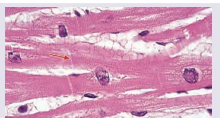
Question 8: Which of the following proteins are not seen in the region marked in the image?
- A. Macula adherens
- B. Fascia adherens
- C. Connexons
- D. Zona occludens (Correct Answer)
Explanation: ***Zona occludens*** - The image shows **cardiac muscle** tissue, and the arrow points to an **intercalated disc**. - Intercalated discs are primarily composed of **fascia adherens**, **maculae adherentes (desmosomes)**, and **gap junctions (connexons)**, but not tight junctions (zona occludens). *Macula adherens* - **Maculae adherentes**, also known as **desmosomes**, are abundant in intercalated discs. - They provide **strong adhesion** between cardiac muscle cells and are crucial for resisting mechanical stress. *Fascia adherens* - **Fascia adherens** are the most extensive type of junction in the transverse portion of the intercalated disc. - They anchor the **actin filaments** of the terminal sarcomeres to the plasma membrane. *Connexions* - **Connexons** are the structural proteins that form **gap junctions**. - Gap junctions in intercalated discs allow for the rapid **passage of ions** and small molecules, facilitating electrical coupling and coordinated contraction.
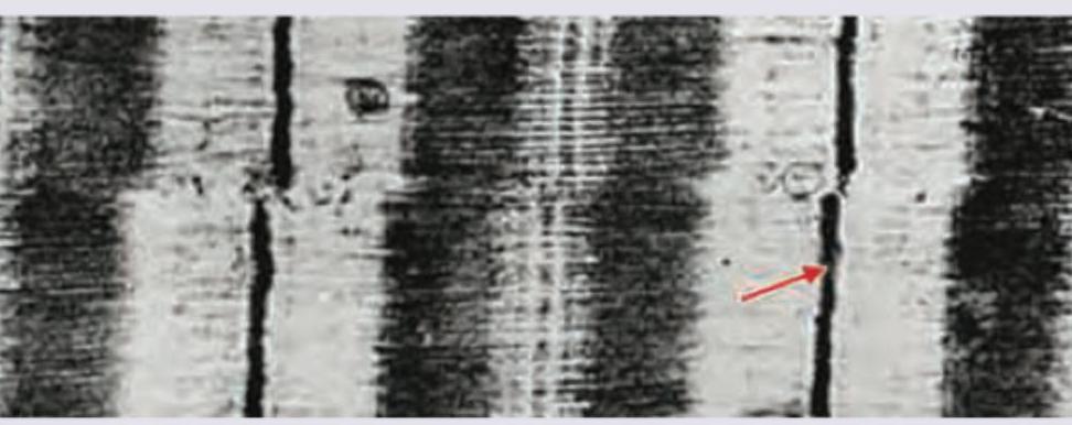
Question 9: Which of the following is the constituent of the marked area in the given electron microscope picture of the muscle?
- A. $\alpha$-actinin (Correct Answer)
- B. Nebulin
- C. Titin
- D. Tropomodulin
Explanation: ***$\alpha$-actinin*** - The image highlights the **Z-disc**, which is primarily composed of **$\alpha$-actinin**. - **$\alpha$-actinin** anchors the **thin filaments (actin)** at the Z-disc and helps maintain the structural integrity of the sarcomere. *Nebulin* - **Nebulin** is a large protein associated with thin filaments, regulating their **length** and contributing to their **stability**, but it is not the main constituent of the Z-disc. - It extends along the entire length of the thin filament, rather than forming the Z-disc itself. *Titin* - **Titin** is the largest known protein, responsible for the **elasticity** of muscle and connecting the Z-disc to the M-line. - While it associates with the Z-disc, it does not constitute the primary structural component of the Z-disc itself. *Tropomodulin* - **Tropomodulin** caps the **pointed (minus) end** of the **actin filaments**, regulating their length and ensuring stability in the sarcomere. - It is located at the ends of the thin filaments, away from the Z-disc.