All SubjectsAnatomy (42)Anesthesiology (16)Biochemistry (24)Community Medicine (55)Dermatology (14)ENT (18)Forensic Medicine (17)General Medicine (2)Internal Medicine (81)Microbiology (32)Obstetrics and Gynecology (59)Ophthalmology (25)Orthopaedics (11)Pathology (44)Pediatrics (40)Pharmacology (29)Physiology (18)Psychiatry (14)Radiology (21)Surgery (45)
Q11
Identify the type of diaphragmatic hernia shown in the X-ray.
Q12
A 70-year-old patient presents with absolute constipation and abdominal distension. The X-ray abdomen is given below. What is the most likely diagnosis?
Q13
Identify the sign given in the image below:
Q14
Which instrument is primarily used to establish pneumoperitoneum in closed laparoscopy technique?
Q15
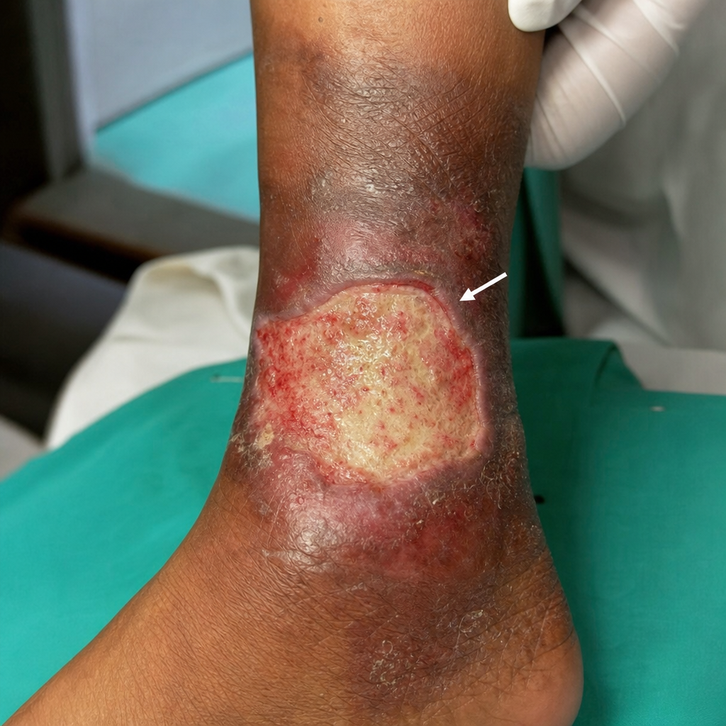
The given image shows an ulcer. Identify the marked structure.
Q16
A 33-year-old male presents with sudden onset acute abdominal pain, constipation for 1 day, persistent hiccups, and occasional vomiting. An abdominal X-ray was performed. Identify the pathology.
Q17
During laparoscopic surgery, which vessel(s) should be specifically avoided during lateral trocar insertion?
Q18
A patient with diffuse severely contaminated peritonitis underwent laparotomy and was left open after surgery. Which of the following might help?
Q19
A patient presents to the emergency department with confusion. On examination, he opens his eyes to pain, shows abnormal flexion to pain, and is disoriented in speech. What is his Glasgow Coma Scale (GCS) score?
Q20
Identify the part of the ulcer indicated by the arrow in the image.