All SubjectsAnatomy (42)Anesthesiology (16)Biochemistry (24)Community Medicine (55)Dermatology (14)ENT (18)Forensic Medicine (17)General Medicine (2)Internal Medicine (81)Microbiology (32)Obstetrics and Gynecology (59)Ophthalmology (25)Orthopaedics (11)Pathology (44)Pediatrics (40)Pharmacology (29)Physiology (18)Psychiatry (14)Radiology (21)Surgery (45)
Q11
Based on the provided MRI images of the knee (A and B), which show a well-defined fluid collection anterior to the patella, what is the most likely diagnosis?
Q12
A patient presented to the OPD with a sudden onset of shortness of breath. Identify the condition with the radiological image given below.
Q13
A patient presents with SOB and fatigue. CXR was done. What is the diagnosis? 
Q14
A patient presents with severe headache. A CT scan of the brain reveals hyperdense areas in the right basal region, marked as 'X'. Which of the following is the most likely diagnosis?
Q15
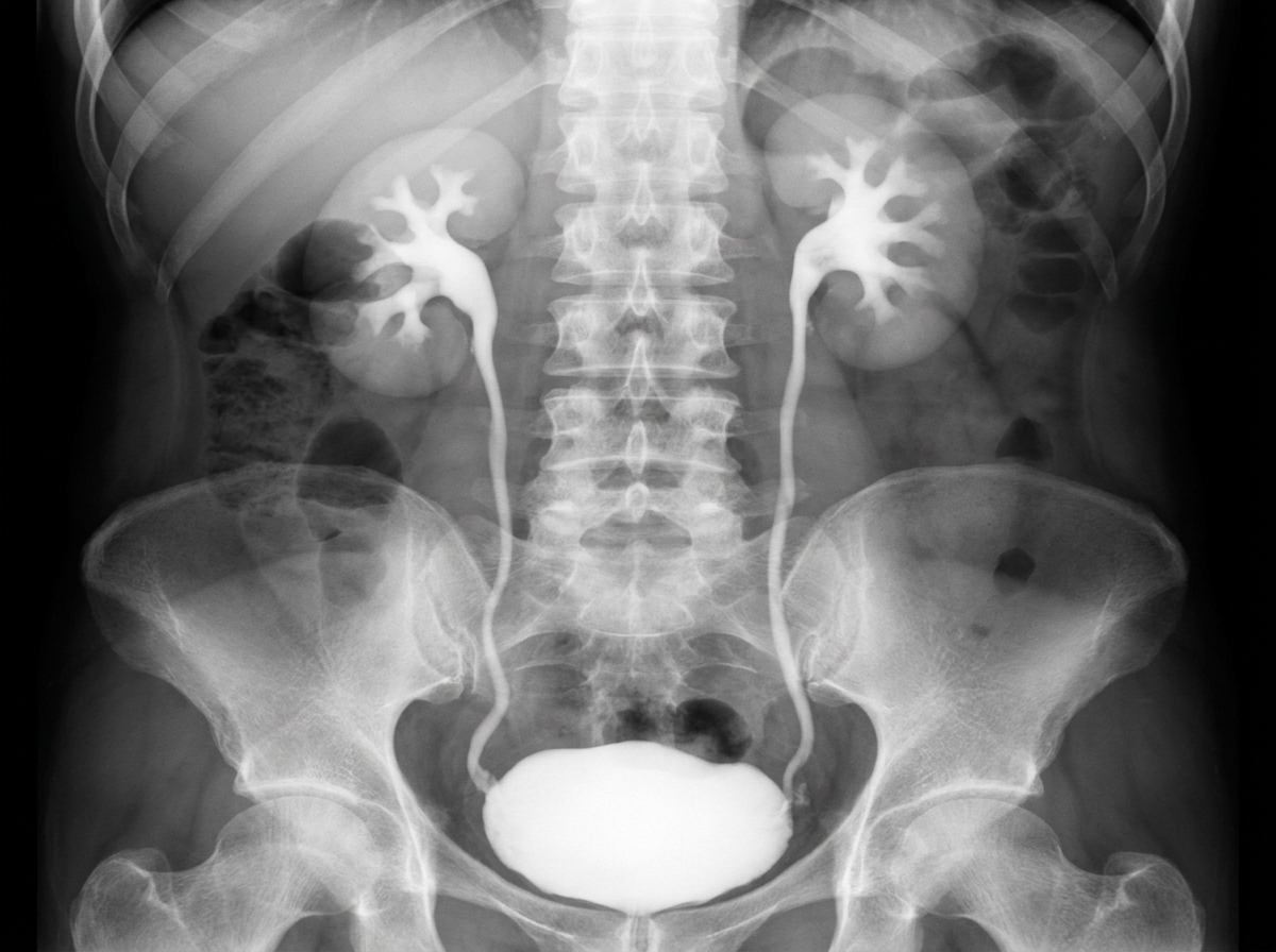
The following radiological image was taken to assess the urinary tract. Identify the investigation shown below.
Q16
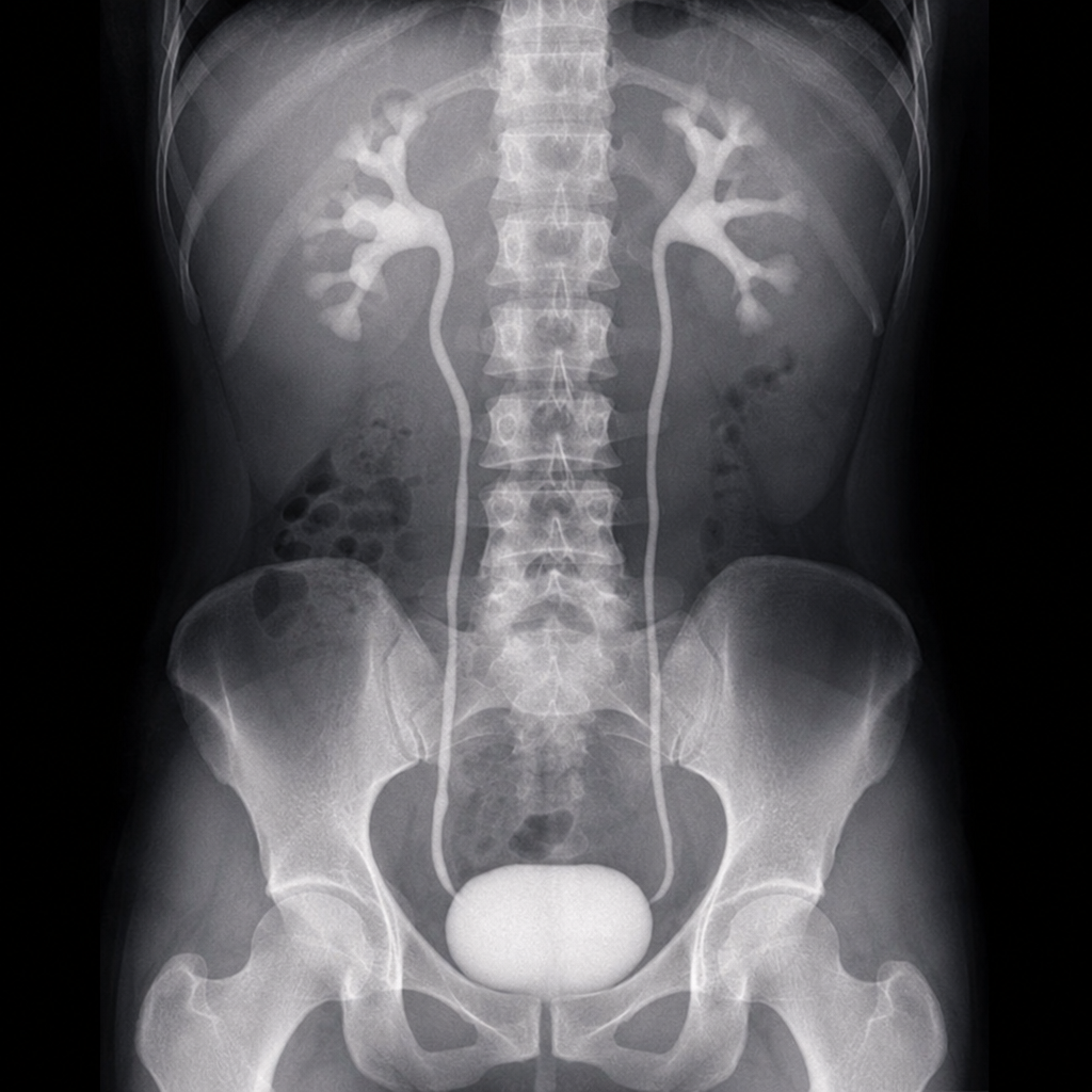
Identify the investigation shown in the image:
Q17
What does a linear accelerator (LINAC) produce in radiation therapy?
Q18
Identify the imaging study shown in the image.
Q19
A patient presents with bilateral hilar lymphadenopathy with eggshell calcification on chest X-ray. What is the most likely diagnosis?
Q20
A 30 year old apparently healthy man who was carrying laxatives and enema apparatus developed abdominal pain at the airport and an x-ray was done which appears as shown below. Which of the following is the likely diagnosis?