All (607)Anatomy (42)Anesthesiology (16)Biochemistry (24)Community Medicine (55)Dermatology (14)ENT (18)Forensic Medicine (17)General Medicine (2)Internal Medicine (81)Microbiology (32)Obstetrics and Gynecology (59)Ophthalmology (25)Orthopaedics (11)Pathology (44)Pediatrics (40)Pharmacology (29)Physiology (18)Psychiatry (14)Radiology (21)Surgery (45)
Q81
What is the likely diagnosis for the given image?
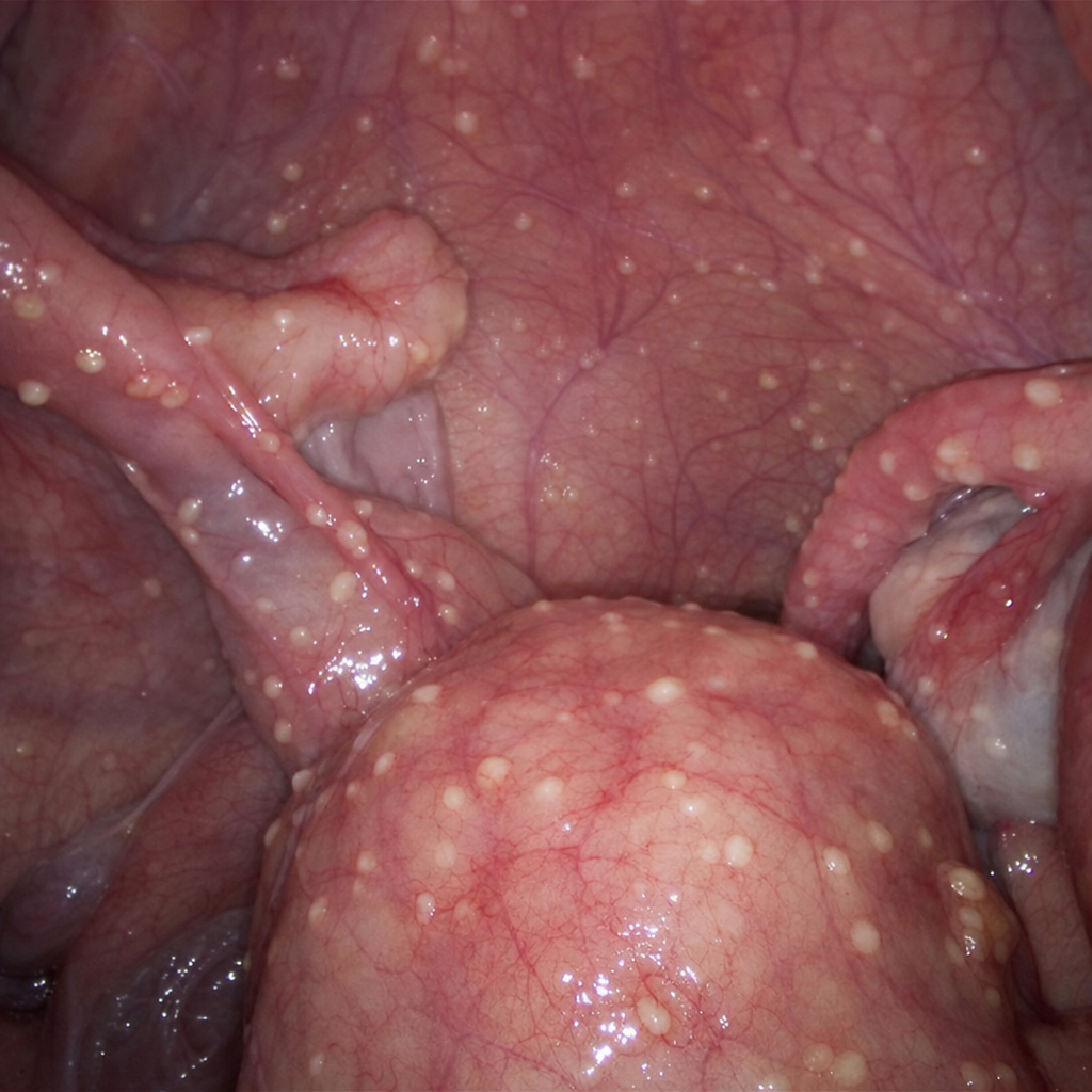
Q82
During laparoscopy, multiple small, yellowish-white tubercles are observed scattered over the pelvic peritoneum and adnexal surfaces. What is the most likely diagnosis?