All (606)Anatomy (42)Anesthesiology (16)Biochemistry (24)Community Medicine (55)Dermatology (14)ENT (18)Forensic Medicine (17)General Medicine (2)Internal Medicine (81)Microbiology (32)Obstetrics and Gynecology (59)Ophthalmology (25)Orthopaedics (11)Pathology (44)Pediatrics (40)Pharmacology (29)Physiology (18)Psychiatry (14)Radiology (21)Surgery (44)
Q381
A patient presents with severe headache. A CT scan of the brain reveals hyperdense areas in the right basal region, marked as 'X'. Which of the following is the most likely diagnosis?
Q382
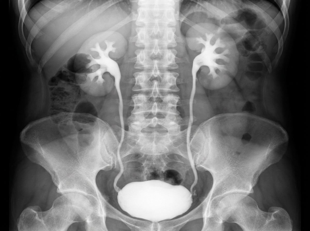
The following radiological image was taken to assess the urinary tract. Identify the investigation shown below.
Q383
Identify the investigation shown in the image:
Q384
What does a linear accelerator (LINAC) produce in radiation therapy?