All (606)Anatomy (42)Anesthesiology (16)Biochemistry (24)Community Medicine (55)Dermatology (14)ENT (18)Forensic Medicine (17)General Medicine (2)Internal Medicine (81)Microbiology (32)Obstetrics and Gynecology (59)Ophthalmology (25)Orthopaedics (11)Pathology (44)Pediatrics (40)Pharmacology (29)Physiology (18)Psychiatry (14)Radiology (21)Surgery (44)
Q271
A patient with diffuse severely contaminated peritonitis underwent laparotomy and was left open after surgery. Which of the following might help?
Q272
A patient presents to the emergency department with confusion. On examination, he opens his eyes to pain, shows abnormal flexion to pain, and is disoriented in speech. What is his Glasgow Coma Scale (GCS) score?
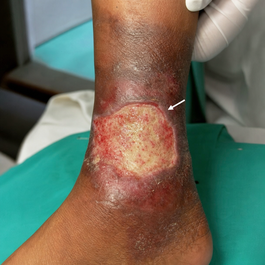
Q273
Identify the part of the ulcer indicated by the arrow in the image.
Q274
A 34-year-old male undergoes an open appendectomy for acute appendicitis. The choice of incision was McBurney's incision. Postoperatively, after a few days, he presents with pain and bulging in the right lower quadrant, which is diagnosed as an indirect inguinal hernia. Which nerve injury during the appendectomy is most likely responsible for this complication?