All SubjectsAnatomy (42)Anesthesiology (16)Biochemistry (24)Community Medicine (55)Dermatology (14)ENT (18)Forensic Medicine (17)General Medicine (2)Internal Medicine (81)Microbiology (32)Obstetrics and Gynecology (59)Ophthalmology (25)Orthopaedics (11)Pathology (44)Pediatrics (40)Pharmacology (29)Physiology (18)Psychiatry (14)Radiology (21)Surgery (44)
Q21
Which antibody is the first to appear and acts fastest during a primary (first) immune response to an infection?
Q22
Chocolate agar is primarily used for the isolation of which of the following organisms?
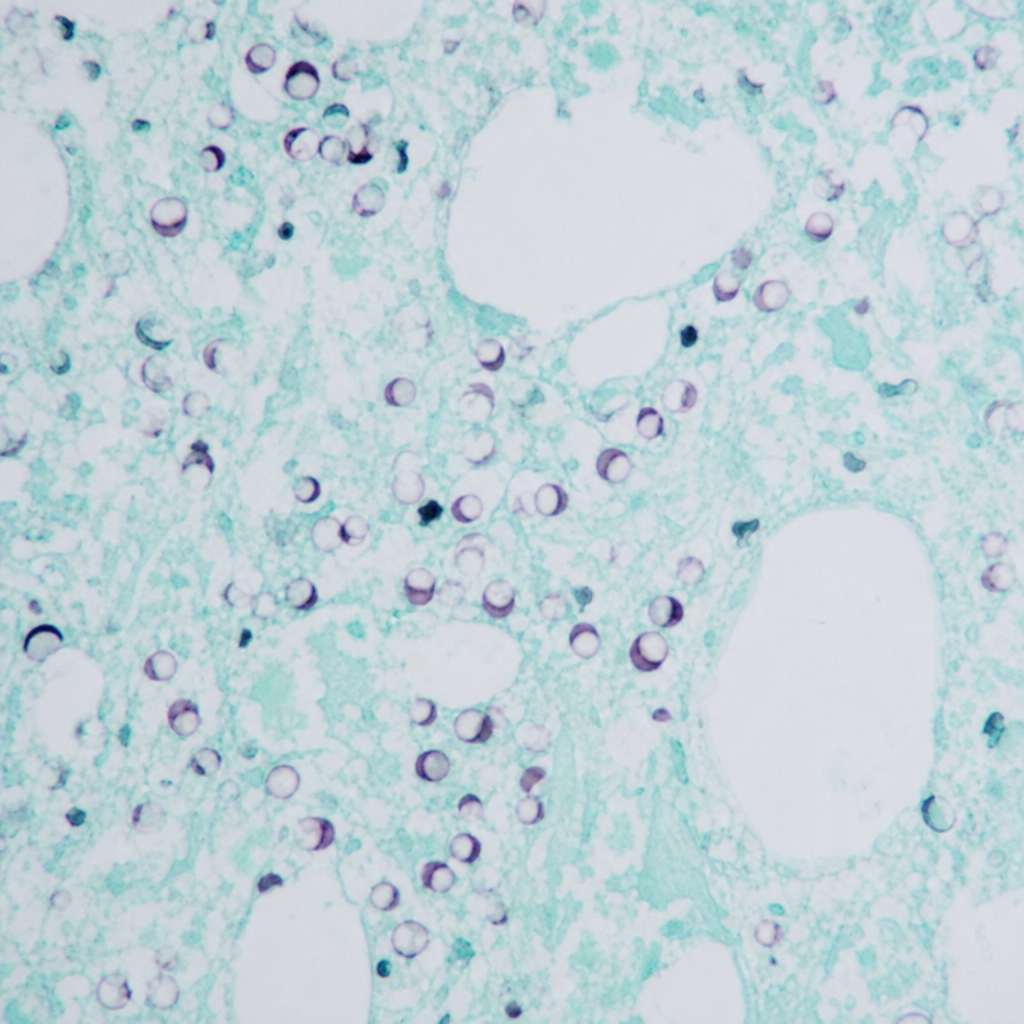
Q23
Identify the opportunistic pathogen demonstrating a 'crushed ping pong ball appearance' on Gomori methenamine stain shown in the image.
Q24
A farmer presents with fever, malaise, and diarrhea. There is a history of exposure to livestock. Which of the following organisms is most likely responsible for the illness?
Q25
A diabetic patient presents with whitish plaques in the oral cavity. A KOH mount confirms the presence of budding yeast with pseudohyphae. Which of the following is the most appropriate culture medium for isolation of this organism?
Q26
A 30-year-old man develops profuse vomiting and diarrhea within 3 hours of consuming pre-packaged salad and milk at a picnic. There is no fever. Which of the following is the most likely causative organism?
Q27
Receptor used for the entry of HIV virus into the host cell is?
Q28
An AIDS-positive patient came with a history of fever, vomiting, and meningismus. Which of the following tests help in the rapid diagnosis of cryptococcal meningitis?
Q29
Which organism is most commonly responsible for gas gangrene?
Q30
A woman was bitten by a rabid dog. The dog developed symptoms and died within a week. What is the best method for post-mortem diagnosis of rabies in the dog?