All (282)Anatomy (15)Anesthesiology (7)Biochemistry (17)Community Medicine (20)Dental (3)Dermatology (2)ENT (7)Forensic Medicine (9)Internal Medicine (36)Microbiology (23)Obstetrics and Gynecology (28)Ophthalmology (13)Orthopaedics (7)Pathology (12)Pediatrics (9)Pharmacology (27)Physiology (11)Psychiatry (7)Radiology (8)Surgery (21)
Q101
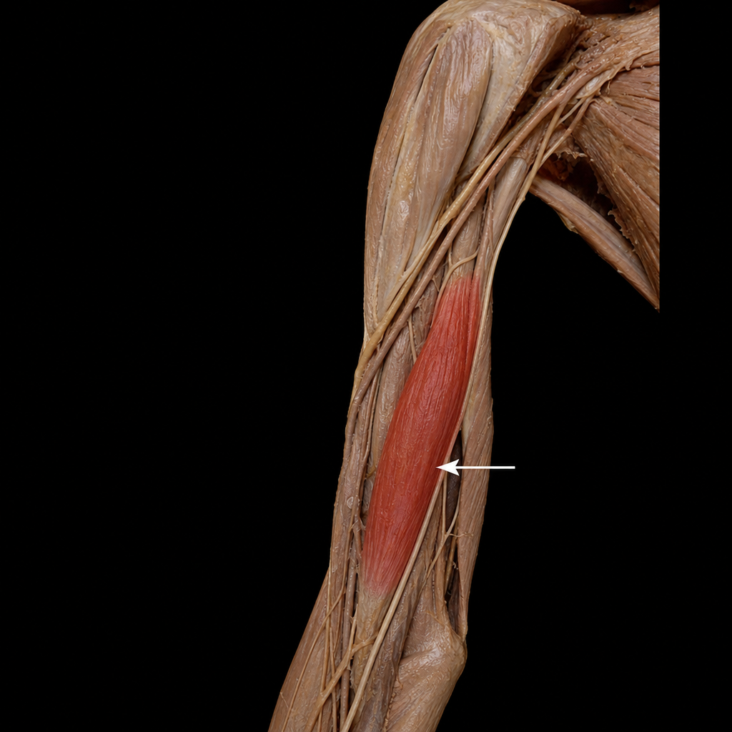
The highlighted muscle is supplied by which nerve?