FMGE 2019 — Orthopaedics
5 Previous Year Questions with Answers & Explanations
Post-menopausal woman, fell down in washroom. What is the most common fracture she may suffer?
Football player with knee injury diagnosed as medial collateral ligament injury. Which structure is most commonly associated with this type of injury?
A 6-year-old child is suspected with supracondylar fracture of right hand, complaining of pain and swelling. X-ray of right elbow was not significant. What is the next best step in this case?
The shown apparatus is used for

An injury to the shown area can lead to fracture of which bone?
FMGE 2019 - Orthopaedics FMGE Practice Questions and MCQs
Question 1: Post-menopausal woman, fell down in washroom. What is the most common fracture she may suffer?
- A. Smith fracture
- B. Colles fracture (Correct Answer)
- C. Monteggia's fracture
- D. Galeazzi fracture
Explanation: ***Colles fracture*** - This fracture commonly occurs in **post-menopausal women** due to **osteoporosis** and typically results from a fall onto an **outstretched hand**. - It involves a **distal radius fracture** with **dorsal displacement** and often radial angulation. *Smith fracture* - A Smith fracture involves a **distal radius fracture** with **volar displacement**, usually caused by a fall onto the back of the hand. - While it can occur in post-menopausal women, it is less common than a Colles fracture in such a scenario. *Monteggia's fracture* - This fracture involves a **fracture of the ulna** with **dislocation of the radial head**. - It usually results from a direct blow to the forearm or a fall with extreme pronation, which is less typical for a simple fall in a post-menopausal woman. *Galeazzi fracture* - A Galeazzi fracture involves a **fracture of the radius** with **dislocation of the distal radioulnar joint (DRUJ)**. - This injury is less common and typically results from a fall onto an outstretched hand with the forearm in pronation, and it is not the most common fracture in this demographic.
Question 2: Football player with knee injury diagnosed as medial collateral ligament injury. Which structure is most commonly associated with this type of injury?
- A. Lateral meniscus
- B. Medial meniscus (Correct Answer)
- C. Anterior cruciate ligament
- D. Posterior Cruciate Ligament
Explanation: ***Medial meniscus*** - The **medial meniscus** is commonly injured in conjunction with the **medial collateral ligament** due to their anatomical proximity and shared role in knee stability. - The MCL is a primary restraint to **valgus stress**, and strong valgus forces that injure the MCL can also transmit stress to the medial meniscus, leading to tears. *Lateral meniscus* - The **lateral meniscus** is less frequently injured alongside the MCL because it is typically more mobile and not directly attached to the MCL. - Injuries to the lateral meniscus are more often associated with **anterior cruciate ligament (ACL) tears** or significant *rotational forces*. *Anterior cruciate ligament* - The **anterior cruciate ligament** is primarily injured by **non-contact pivoting** or **hyperextension injuries**, and while it can be part of the "unhappy triad" (along with MCL and medial meniscus tears), an isolated MCL injury does not most commonly implicate it. - ACL tears lead to **anterior instability** of the tibia relative to the femur, which is a different biomechanical mechanism than an isolated MCL injury. *Posterior Cruciate Ligament* - The **posterior cruciate ligament** is injured by a direct blow to the anterior tibia while the knee is flexed or during a dashboard injury, leading to **posterior instability**. - Its injury mechanism is distinct from that of the MCL, which is primarily due to **valgus stress**.
Question 3: A 6-year-old child is suspected with supracondylar fracture of right hand, complaining of pain and swelling. X-ray of right elbow was not significant. What is the next best step in this case?
- A. Cast
- B. Closed reduction with K wire fixation
- C. Compare with X-ray of left hand (Correct Answer)
- D. Closed reduction and slab
Explanation: ***Compare with X-ray of left hand*** - In pediatric elbow injuries, a seemingly **normal X-ray** in the presence of strong clinical suspicion (pain, swelling, suspected supracondylar fracture) often warrants a comparison view of the contralateral unaffected limb. - This helps identify subtle findings like **epiphyseal separations** or **minimally displaced fractures** that might otherwise be missed due to the developing osseous structures in children. *Cast* - Applying a cast without definitive diagnosis or clear radiographic evidence of a fracture can lead to **unnecessary immobilization** and potential complications if no fracture is present, or inadequate treatment if a specific type of fracture requires reduction. - While immobilization is appropriate for confirmed fractures, it's not the **initial diagnostic step** when X-rays are inconclusive. *Closed reduction with K wire fixation* - This is an **invasive procedure** reserved for **displaced or unstable fractures** after a clear diagnosis has been established. - Performing this without a confirmed and characterized fracture is inappropriate and carries risks of **iatrogenic injury** and complications. *Closed reduction and slab* - Similar to casting, this is a treatment for **confirmed fractures**, typically for acute, stable, or minimally displaced fractures that can be managed non-surgically after a reduction. - It is not a diagnostic step and should not be performed when initial imaging is **inconclusive** and the exact nature of the injury is unknown.
Question 4: The shown apparatus is used for
- A. Ankle knee stabilizer
- B. Thomas splint
- C. Knee brace
- D. Patella tendon bearing brace (Correct Answer)
Explanation: ***Patella tendon bearing brace*** - This orthotic device is designed to **transfer weight-bearing load through the patella tendon**, reducing stress on the lower extremity during ambulation. - It features a **molded cuff** that fits snugly below the patella and distributes weight through the **patellar tendon bearing area**, commonly used in **prosthetic applications** and **below-knee amputees**. *Ankle knee stabilizer* - This device provides **combined support to both ankle and knee joints** simultaneously, typically used for **multi-joint injuries** or instability. - It features **dual bracing systems** with straps and supports extending from ankle to knee, unlike the focused patellar tendon bearing design. *Thomas splint* - A **rigid metal-framed splint** used primarily for **femur fracture stabilization** and maintaining **skeletal traction** in emergency situations. - It consists of a **ring that fits around the upper thigh** with extending metal bars, designed for **fracture immobilization** rather than weight distribution. *Knee brace* - A general **knee joint support device** used for **ligament injuries**, **post-surgical recovery**, or **osteoarthritis management**. - Available in various forms (**sleeve, hinged, or wraparound designs**) but lacks the specific **weight-bearing transfer mechanism** of a patella tendon bearing brace.
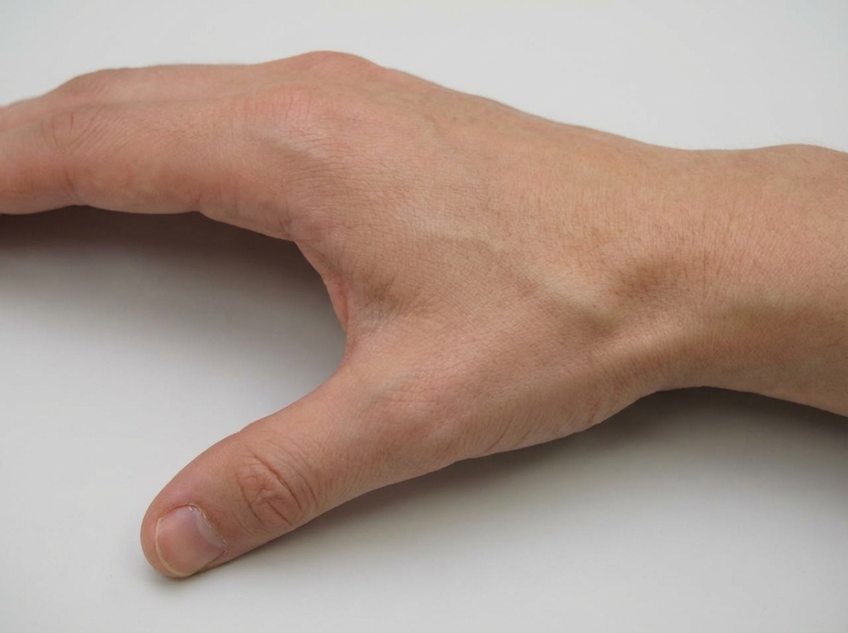
Question 5: An injury to the shown area can lead to fracture of which bone?
- A. Lunate
- B. Scaphoid (Correct Answer)
- C. Hamate
- D. Trapezium
Explanation: ***Scaphoid*** - The **anatomical snuffbox** is the most vulnerable area for scaphoid fractures, typically occurring after a **fall on outstretched hand (FOOSH)** injury. - **Tenderness in the anatomical snuffbox** and pain with **radial deviation** of the wrist are classic clinical findings of scaphoid fracture. *Lunate* - Lunate fractures are much **rarer** and typically occur with **high-energy trauma** or **perilunate dislocations**. - The lunate is **not directly palpable** in the anatomical snuffbox area, making it less susceptible to injury from this mechanism. *Hamate* - Hamate fractures commonly occur from **direct trauma** to the **hypothenar eminence** or **hook of hamate**. - The hamate is located on the **ulnar side** of the wrist and is not anatomically related to the snuffbox area. *Trapezium* - Trapezium fractures are typically associated with **Bennett's fracture** involving the **first metacarpal base**. - Located at the **base of the thumb**, the trapezium is not directly involved in anatomical snuffbox injuries.