All SubjectsAnatomy (15)Anesthesiology (7)Biochemistry (17)Community Medicine (20)Dental (3)Dermatology (2)ENT (7)Forensic Medicine (9)Internal Medicine (36)Microbiology (23)Obstetrics and Gynecology (28)Ophthalmology (13)Orthopaedics (7)Pathology (12)Pediatrics (9)Pharmacology (27)Physiology (11)Psychiatry (7)Radiology (8)Surgery (21)
Q11
Trigone of bladder is derived from?
Q12
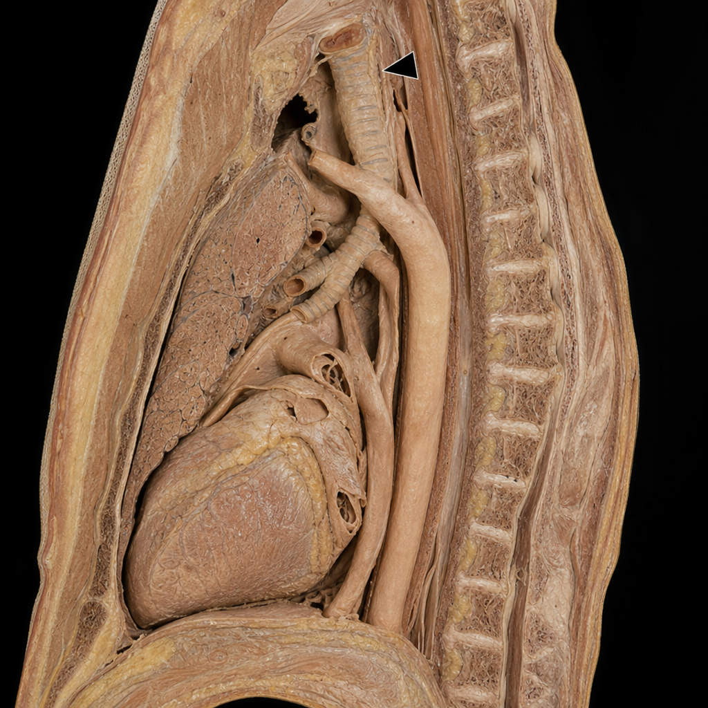
In the shown lateral view, the structure marked with arrowhead is?
Q13
Visual loss due to cerebral degeneration is related to which artery?
Q14
Adam's apple in males is formed by the
Q15
Avascular necrosis of the femoral head most commonly occurs due to disruption of which of the following arteries?