

Kidney Gross Anatomy - Bean Scene Investigation
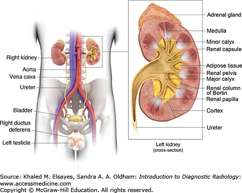
- Location: Retroperitoneal organs at T12-L3 vertebral levels. The right kidney is slightly lower than the left due to the liver's position.
- Coverings (Outermost to Innermost):
- Pararenal fat: Outermost layer of extraperitoneal fat.
- Renal fascia (Gerota's): Encloses kidney and adrenal gland.
- Perirenal fat: Cushions the kidney.
- Fibrous capsule: Adheres directly to the kidney surface.
- Hilum (Anterior to Posterior): Renal Vein → Renal Artery → Renal Pelvis.
- 📌 Mnemonic: VAP.
- Parenchyma:
- Cortex: Outer granular region containing glomeruli & convoluted tubules.
- Medulla: Inner region with 8-18 renal pyramids.
- Urine Flow: Papilla of pyramid → Minor Calyx → Major Calyx → Renal Pelvis → Ureter.
⭐ The left renal vein passes between the superior mesenteric artery (SMA) and the abdominal aorta, creating a risk for compression (Nutcracker Syndrome), which can cause hematuria and flank pain.
Renal Vasculature - Blood Flow Blowout
- Arterial Supply: Renal arteries arise from the abdominal aorta, branching into segmental arteries.
- Segmental arteries are end-arteries; occlusion leads to infarction.
- Path: Aorta → Renal a. → Segmental a. → Interlobar a. → Arcuate a. → Interlobular a. → Afferent arteriole.
- Venous Drainage: Follows arterial path in reverse, ultimately draining into the Inferior Vena Cava (IVC).
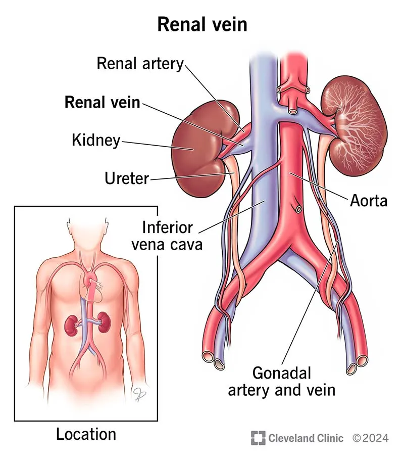
⭐ Left Renal Vein: The longer left renal vein passes between the aorta and SMA. It receives the left suprarenal and left gonadal (testicular/ovarian) veins, unlike the right side. This can lead to "Nutcracker Syndrome."
Suprarenal Glands - Gland Stand
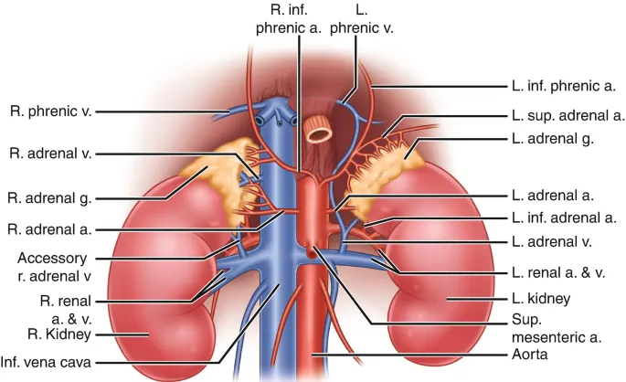
- Arterial Supply: Rich and redundant from three sources:
- Superior suprarenal a.: from inferior phrenic a.
- Middle suprarenal a.: from abdominal aorta.
- Inferior suprarenal a.: from renal a.
- Venous Drainage: A single vein per gland.
| Feature | Cortex | Medulla |
|---|---|---|
| Embryology | Mesoderm | Neural Crest Cells |
| Function | 📌 G-F-R for layers; Salt-Sugar-Sex for products | Synthesizes & secretes catecholamines |
| Layers | Glomerulosa, Fasciculata, Reticularis | Chromaffin cells |
| Products | Aldosterone, Cortisol, Androgens | Epinephrine, Norepinephrine |
- The left renal vein, unlike the right, receives the left suprarenal and left gonadal veins before draining into the IVC.
- Nutcracker syndrome involves compression of the left renal vein between the aorta and the superior mesenteric artery.
- Ureters have three physiological constrictions where stones often lodge: ureteropelvic junction, crossing the common iliac artery, and the ureterovesical junction.
- The adrenal cortex (mesoderm) secretes steroids, while the medulla (neural crest) secretes catecholamines.
- A horseshoe kidney gets trapped under the inferior mesenteric artery (IMA) during ascent.
Unlock the full lesson and continue reading
Signup to continue reading this lesson and unlimited access questions, flashcards, AI notes, and more