

Temporal Fossa - Temple Territory
- Boundaries:
- Superior & Posterior: Superior temporal line.
- Anterior: Frontal & zygomatic bones.
- Lateral: Zygomatic arch.
- Inferior: Infratemporal crest (greater wing of sphenoid).
- Floor: Pterion (junction of Frontal, Parietal, Temporal, Sphenoid bones 📌FPTS).
- Roof: Temporal fascia (superficial layer).
- Contents:
- Temporalis muscle.
- Temporal fascia (deep layer, investing temporalis m.).
- Deep temporal nerves (anterior & posterior branches of mandibular nerve V3).
- Deep temporal arteries (anterior & posterior; from maxillary artery) & accompanying veins.

⭐ Pterion: key floor landmark; overlies middle meningeal artery. Fracture risks epidural hematoma.
Infratemporal Fossa - Deep Skull Base
- Irregular space inferior & deep to zygomatic arch.
- Boundaries:
- Lateral: Mandibular ramus.
- Medial: Lat. pterygoid plate.
- Anterior: Post. maxilla.
- Posterior: Tympanic plate, styloid/mastoid.
- Roof: Sphenoid greater wing, squamous temporal.
- Floor: Open to neck.
- Communications & Key Foramina:
- Foramen Ovale: V3, Acc. meningeal a., Lesser petrosal n., Emissary v. (📌 OVALE)
- Foramen Spinosum: Middle meningeal a./v., nervus spinosus.
- Pterygomaxillary Fissure: To pterygopalatine fossa.
- Contents Overview:
- Muscles: Medial/Lateral pterygoids, temporalis (inf. part).
- Nerves: V3 branches, chorda tympani, otic ganglion.
- Vessels: Maxillary artery (1st/2nd parts), pterygoid venous plexus.
⭐ Foramen Ovale transmits: Mandibular Nerve (V3), Accessory meningeal artery, Lesser petrosal nerve, Emissary vein.
Muscles & Mandibular Nerve - Chew & Feel Central
Muscles of Mastication: All innervated by Mandibular Nerve (CN V3).
| Muscle | O | I | A |
|---|---|---|---|
| Masseter | Zygomatic arch | Mandibular ramus | Elevate, protract |
| Temporalis | Temporal fossa | Coronoid proc. | Elevate, retract |
| Medial Pterygoid | Med. pterygoid plate | Med. mandibular ramus | Elevate, protract, side-to-side |
| Lateral Pterygoid | Lat. pterygoid plate | Mandibular neck, TMJ disc | Protract, depress, side-to-side |
Mandibular Nerve (CN V3): Mixed; exits Foramen Ovale.
- 📌 Mnemonic (Branches): "Bad Apples Look Incredibly Mouthwatering" (Buccal, Auriculotemporal, Lingual, Inferior Alveolar, Motor branches).
- From Trunk: N. to medial pterygoid, Meningeal br.
- Anterior Div (motor focus): Nerves to masseter, temporalis, lat. pterygoid; Buccal n. (sensory).
- **Posterior Div (sensory focus):
- Auriculotemporal n. (sensory; parotid secretomotor via otic ganglion).
- Lingual n. (sensory ant. 2/3 tongue; +chorda tympani CN VII).
- Inferior alveolar n. (sensory lower teeth; N. to mylohyoid motor; Mental n. terminal).
Loading diagram…
Otic Ganglion:
- Location: Infratemporal fossa, medial to V3, inferior to foramen ovale.
- Function: Parasympathetic; relays lesser petrosal n. (CN IX) fibers to parotid gland via auriculotemporal n. (secretomotor).
⭐ Lateral pterygoid is the only muscle of mastication that primarily opens (depresses) the jaw.
Maxillary Artery & TMJ - Blood, Bites, Breaks
- Maxillary Artery: From ECA.
- 1st (Mandibular): Middle Meningeal A. (→ foramen spinosum), Inferior Alveolar A.
- 2nd (Pterygoid): Muscular branches (to masticatory muscles).
- 3rd (Pterygopalatine): Sphenopalatine A. (terminal), Infraorbital A., Post. Sup. Alveolar A.
Loading diagram…
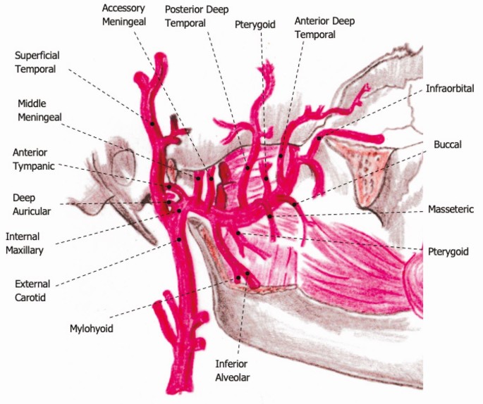
> ⭐ The Middle Meningeal Artery, branch of 1st part maxillary artery, enters via foramen spinosum. Rupture (e.g. pterion fracture) causes extradural hematoma.
-
Pterygoid Venous Plexus:
- Location: Around lateral pterygoid muscle.
- Drains: To maxillary vein.
- Connects: Cavernous sinus, facial vein.
- Clinical: Infection spread to cavernous sinus.
-
Temporomandibular Joint (TMJ):
- Type: Synovial (condylar & hinge).
- Articular Surfaces: Mandibular condyle, temporal bone's articular fossa & tubercle.
- Articular Disc: Divides joint; upper (gliding), lower (hinge).
- Ligaments: Lateral, Sphenomandibular, Stylomandibular.
- Movements & Muscles:
Movement Primary Muscles Elevation Masseter, Temporalis, Medial pterygoid Depression Lateral pterygoid, Suprahyoids Protrusion Lateral pterygoid, Medial pterygoid Retrusion Temporalis (post.), Masseter (deep) Lateral Contra Pterygoids, Ipsi Temporalis
-
Key Clinicals:
- TMJ Dislocation: Anteriorly; mandible open.
- Frey's Syndrome: Gustatory sweating (auriculotemporal n. injury).
- Trismus: Limited mouth opening (pterygoid spasm).
High‑Yield Points - ⚡ Biggest Takeaways
- Muscles of mastication: Innervated by Mandibular N. (V3). Lateral pterygoid opens jaw.
- Mandibular N. (V3): Exits foramen ovale. Middle Meningeal A. (maxillary branch) through foramen spinosum.
- Otic Ganglion: Medial to V3; relays CN IX parasympathetics to parotid via auriculotemporal N.
- Chorda Tympani (CN VII): Joins Lingual N. (V3); taste (ant. 2/3 tongue) & secretomotor.
- Inferior Alveolar N.: V3 branch, supplies lower teeth, enters mandibular foramen.
- Pterygoid Venous Plexus: Risk for infection spread to cavernous sinus_
Unlock the full lesson and continue reading
Signup to continue reading this lesson and unlimited access questions, flashcards, AI notes, and more