

Neck Mass Map - Region & Reason
- Midline Structures:
- Thyroglossal duct cyst: congenital, moves with tongue protrusion/swallowing.
- Thyroid: goiter, nodule, carcinoma; often requires USG/FNAC.
- Dermoid cyst: congenital, doughy consistency, may contain adnexal structures.
- Submental lymphadenopathy: often due to dental/oral infections.
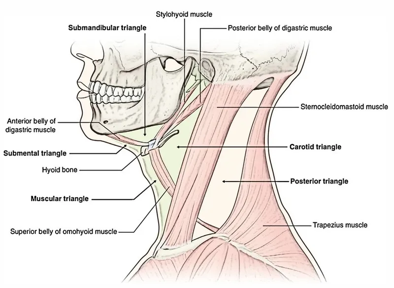
- Anterior Triangle:
- Submandibular: Salivary gland (sialadenitis, tumor), regional lymph nodes.
- Carotid region: 2nd Branchial cleft cyst (anterior to SCM), carotid body tumor (pulsatile), cervical lymphadenopathy.
- Posterior Triangle:
- Lymphadenopathy: commonest; metastatic (SCC), lymphoma, TB (scrofula).
- Cystic hygroma (lymphangioma): congenital, soft, transilluminates.
- Lipoma, neurogenic tumors (schwannoma, neurofibroma).
- Supraclavicular Fossa:
- Lymphadenopathy: high suspicion of malignancy (e.g., lung, GI).
- Cystic hygroma, lipoma.
⭐ Virchow's node (left supraclavicular) strongly suggests metastatic gastric cancer; part of Troisier's sign.
The Patient Story - Clues & Feels
- History:
- Onset/Duration: Acute (inflam/infect) vs. Chronic (neoplasm/congenital).
- Pain: Painful (inflam) vs. Painless (neoplasm/congenital).
- Red Flags: Hoarseness, dysphagia, weight loss, night sweats.
- Risk Factors: Smoking, alcohol, radiation, family Hx cancer.
- Systemic: Fever (infection), thyroid symptoms.
- Examination ("Feels"):
- Location: Midline, lateral; specific triangles.
- Consistency: Soft, cystic, firm, rubbery, hard (malignancy).
- Mobility: Mobile (benign) vs. Fixed (malignancy).
- Tenderness: Suggests inflammation.
- Special Signs:
- Swallowing movement: Thyroid, thyroglossal cyst.
- Tongue protrusion movement: Thyroglossal cyst. 📌 (Sistrunk's sign)
⭐ A persistent, firm, enlarging neck mass in an adult, especially >40 years with smoking history, is highly suspicious for malignancy.
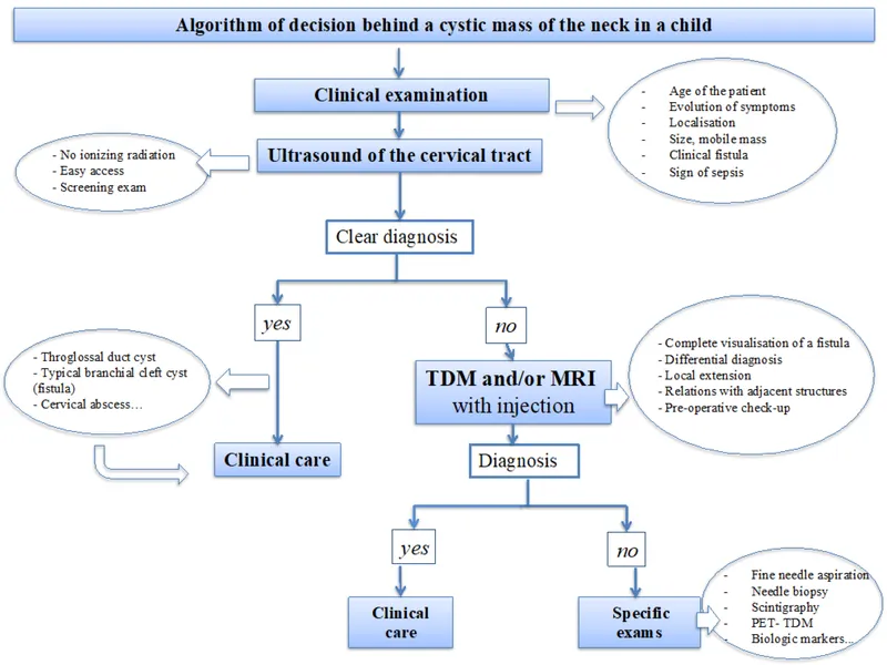
Investigation Arsenal - Scan & Sample
- Initial Scan:
- Ultrasound (USG): First-line. Differentiates cystic vs. solid. Guides FNA.

- Ultrasound (USG): First-line. Differentiates cystic vs. solid. Guides FNA.
- Advanced Scans (Staging & Extent):
- CECT Neck: Defines extent, lymph node status (size >1.5cm, necrosis, ECE).
- MRI: Superior for soft tissue (e.g., parapharyngeal, perineural spread).
- PET-CT: For unknown primary, staging.
- Tissue Sampling:
- FNAC (Fine Needle Aspiration Cytology): Gold standard initial diagnosis. USG-guided for ↑accuracy.
⭐ A negative FNAC in a clinically suspicious node (especially for lymphoma) warrants further investigation, often an excisional biopsy.
- Core Needle Biopsy (CNB): For suspected lymphoma if FNAC non-diagnostic (provides architecture).
- Excisional Biopsy: Definitive diagnosis if other methods fail (esp. lymphoma). Avoid incisional if malignancy suspected (seeding risk).
- FNAC (Fine Needle Aspiration Cytology): Gold standard initial diagnosis. USG-guided for ↑accuracy.
Common Culprits - Rogues' Gallery
-
Thyroglossal Duct Cyst (TGDC)
- Midline (or just off-midline), painless, moves with tongue protrusion & swallowing.
- Embryological remnant.
- Treatment: Sistrunk procedure (excision of cyst, duct, central hyoid).
-
Branchial Cleft Cyst
- Smooth, non-tender, fluctuant mass on lateral neck, anterior to SCM.
- Usually 2nd arch origin.
- Can get infected.
-
Lymphadenopathy (LAD)
- Reactive: Tender, mobile (infection).
- Malignant: Hard, fixed (metastasis, lymphoma).
- Tuberculous (Scrofula): Matted nodes, posterior triangle; cold abscess. 📌 "King's evil".
-
Dermoid & Epidermoid Cysts
- Midline, slow-growing, doughy. Contain keratin/skin adnexa.
-
Lipoma
- Soft, lobulated, mobile, subcutaneous. "Slip sign" positive. Benign.
-
Carotid Body Tumor (Paraganglioma)
- Pulsatile mass at carotid bifurcation.
- Mobile side-to-side, not vertically (Fontaine's sign).
- "Lyre sign" on angiography.
⭐ Thyroglossal duct cysts, the most common congenital midline neck mass, characteristically move upwards with tongue protrusion.
High‑Yield Points - ⚡ Biggest Takeaways
- Persistent neck mass > 2 weeks warrants investigation, especially in adults.
- FNAC is the primary diagnostic tool for most palpable neck masses.
- Midline masses (children): often thyroglossal duct cysts (move with tongue); Lateral masses (children): branchial cleft cysts or lymphadenopathy.
- Adults > 40: firm, fixed, painless lateral neck mass is metastatic SCC until proven otherwise.
- Left supraclavicular mass (Virchow's node) strongly suggests infraclavicular malignancy (gastric, lung, lymphoma).
Unlock the full lesson and continue reading
Signup to continue reading this lesson and unlimited access questions, flashcards, AI notes, and more