

IOFB Definition & Types - The Unwanted Guest
An Intraocular Foreign Body (IOFB) is an object lodged within the eye.
- Definition:
- Penetrating: Entry wound, no exit.
- Perforating: Entry and exit wounds.
- Causes: Hammering (metal-on-metal), occupational hazards.
- Epidemiology: Young males. >50% of open globe injuries may have an IOFB.
| Type | Examples | Reactivity |
|---|---|---|
| Metallic | Iron, Steel, Copper | Reactive (Fe, Cu 📌 "FeCu are foes") |
| Non-metallic | Glass, Stone, Plastic | Generally Inert |
| Organic | Wood, Thorn | Highly Reactive, ↑Infection risk |
| %%{init: {'flowchart': {'htmlLabels': true}}}%% | ||
| flowchart TD |
Start["👁️ Suspected OGI
• Suspect IOFB• Emergent triage"]
Life["⚠️ Life Threats
• Treat concurrents• Trauma protocols"]
Exam["🩺 Eye Exam
• No IOP check• Apply eye shield"]
Meds["💊 Systemic Tx
• IV Antibiotics• Tetanus shot"]
CT["🔬 Orbit CT
• Without contrast• Confirm IOFB"]
Globe["🩹 Globe Closure
• Proceed to repair• Surgical entry"]
Immed["⚡ Immediate removal
• Organic or Toxic• Stable patient"]
Delay["⏳ Delayed removal
• Severe edema• Unstable patient"]
PPV["🔬 Small Gauge PPV
• 6mm infusion• AC infusion"]
Lens["👁️ Lensectomy
• Lens involvement• Remove crystal"]
Vit["✂️ Vitrectomy
• Mobilize IOFB• Biopsy/Cultures"]
Remove["🧲 IOFB Removal
• Exit strategy• Instrumentation"]
Repair["🩺 Retina Repair
• 360 Examination• Tamponade PRN"]
Finish["✅ Final Steps
• Gas or Silicone• IVT Antibiotics"]
Start --> Life Life --> Exam Start --> Exam Exam --> Meds Meds --> CT CT --> Globe Globe -->|Organic| Immed Globe -->|Unstable| Delay Immed --> PPV Delay --> PPV PPV -->|Lens damage| Lens Lens --> Vit PPV --> Vit Vit --> Remove Remove --> Repair Repair --> Finish
style Start fill:#FDF4F3, stroke:#FCE6E4, stroke-width:1.5px, rx:12, ry:12, color:#B91C1C style Life fill:#FDF4F3, stroke:#FCE6E4, stroke-width:1.5px, rx:12, ry:12, color:#B91C1C style Exam fill:#FFF7ED, stroke:#FFEED5, stroke-width:1.5px, rx:12, ry:12, color:#C2410C style Meds fill:#F1FCF5, stroke:#BEF4D8, stroke-width:1.5px, rx:12, ry:12, color:#166534 style CT fill:#FFF7ED, stroke:#FFEED5, stroke-width:1.5px, rx:12, ry:12, color:#C2410C style Globe fill:#F7F5FD, stroke:#F0EDFA, stroke-width:1.5px, rx:12, ry:12, color:#6B21A8 style Immed fill:#FEF8EC, stroke:#FBECCA, stroke-width:1.5px, rx:12, ry:12, color:#854D0E style Delay fill:#FEF8EC, stroke:#FBECCA, stroke-width:1.5px, rx:12, ry:12, color:#854D0E style PPV fill:#F1FCF5, stroke:#BEF4D8, stroke-width:1.5px, rx:12, ry:12, color:#166534 style Lens fill:#F7F5FD, stroke:#F0EDFA, stroke-width:1.5px, rx:12, ry:12, color:#6B21A8 style Vit fill:#F1FCF5, stroke:#BEF4D8, stroke-width:1.5px, rx:12, ry:12, color:#166534 style Remove fill:#F1FCF5, stroke:#BEF4D8, stroke-width:1.5px, rx:12, ry:12, color:#166534 style Repair fill:#F7F5FD, stroke:#F0EDFA, stroke-width:1.5px, rx:12, ry:12, color:#6B21A8 style Finish fill:#F1FCF5, stroke:#BEF4D8, stroke-width:1.5px, rx:12, ry:12, color:#166534
> ⭐ Most common IOFBs are metallic, often from hammering metal-on-metal.
## IOFB Diagnosis - Spotting the Intruder
Key diagnostic steps involve a thorough history, clinical examination, and appropriate imaging.
* **History**
- Mechanism: High-speed projectile (e.g., hammering, explosion, grinding)?
- Material: Suspected type (metallic, glass, organic)?
* **Symptoms**
- Pain (may be minimal), ↓ vision, floaters, photophobia.
* **Signs**
- Entry wound (corneal/scleral): **Seidel's test positive** if aqueous leak present.
- Anterior segment: Hyphema, iris defect (e.g., transillumination, iridodialysis).
- Lens: Focal cataract.
- Posterior segment: Vitreous hemorrhage, cells.
**Imaging Modalities for IOFB Detection & Localization:**
| Imaging | Key Features |
|-----------------|-------------------------------------------------------------------------------------------------------------|
| X-ray (AP/Lat) | Detects radio-opaque FBs; poor localization. Bone-free views helpful. |
| Ultrasound (B-scan) | Dynamic; good for posterior segment, FB detection/location, especially with opaque media. |
| CT Scan (orbital) | **Gold standard** for metallic IOFB; precise localization. Thin non-contrast axial & coronal cuts. |
| MRI | ⚠️ **Contraindicated** if metallic IOFB suspected (risk of movement/heating). Useful for non-metallic FBs. |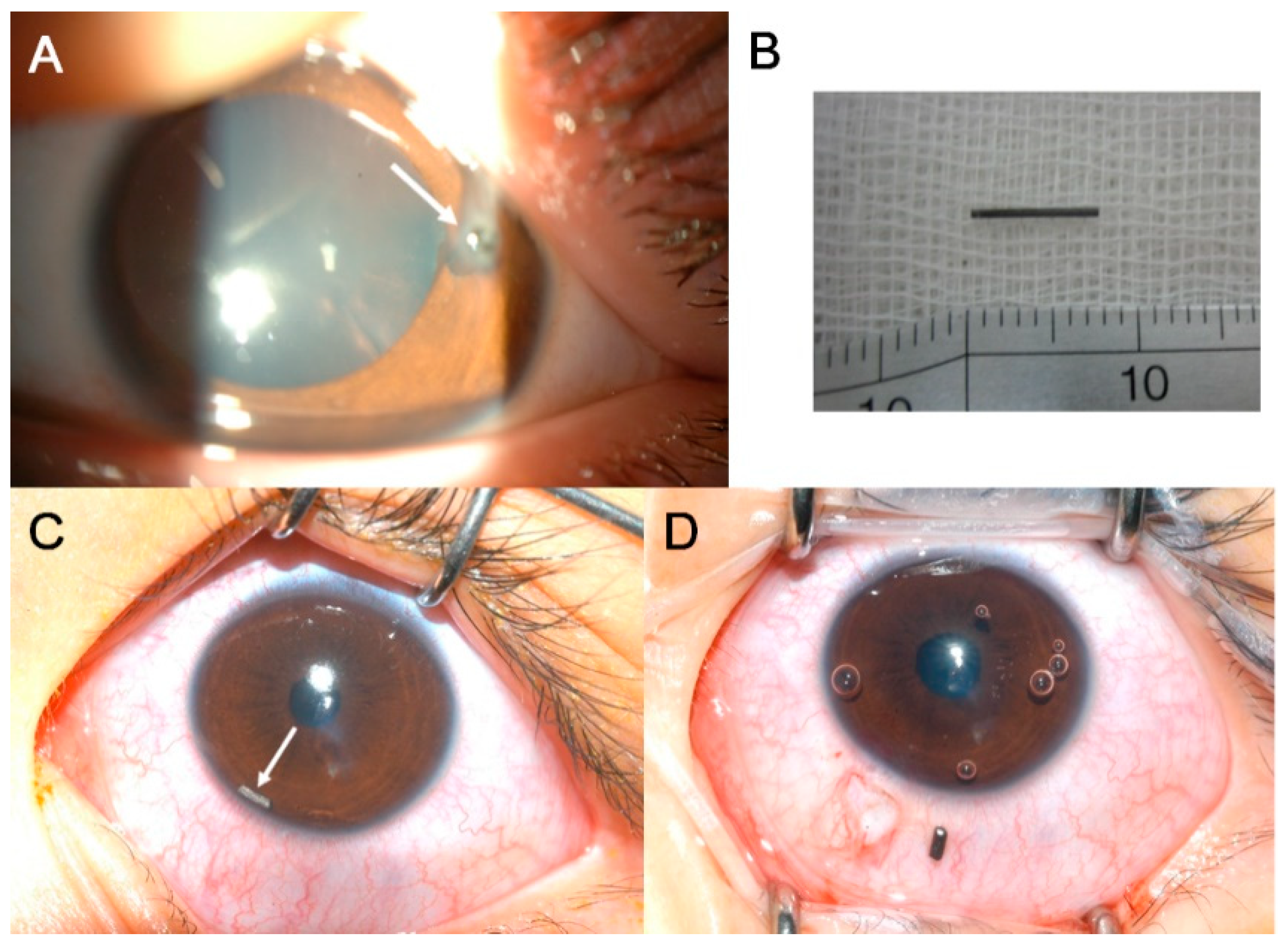
> ⭐ Non-contrast CT scan (thin axial and coronal slices) is the investigation of choice for suspected IOFB due to its high sensitivity and specificity for metallic FBs and precise localization ability.
## IOFB Management - Damage Control Tactics
**Initial Management (Pre-operative):**
* Protect the eye: Rigid eye shield (NO patching).
* NPO (Nil Per Oral): In anticipation of surgery.
* Systemic Prophylaxis:
- Tetanus toxoid (IM/IV) as per status.
- Broad-spectrum IV antibiotics (e.g., Vancomycin + Ceftazidime).
* Topical broad-spectrum antibiotics (e.g., Moxifloxacin 0.5% q1h).
* Control pain & nausea: Analgesics (e.g., Paracetamol) & Antiemetics (e.g., Ondansetron).
**Surgical Management:**
* **Timing:** Urgent, ideally within **24-72 hours** of injury (↓ endophthalmitis risk).
* **Anesthesia:** General Anesthesia (GA) preferred for controlled environment.
**Surgical Approach Decision:**
```mermaid
%%{init: {'flowchart': {'htmlLabels': true}}}%%
flowchart TD
Start["<b>👁️ IOFB Confirmed</b><br><span style='display:block; text-align:left; color:#555'>• Intraocular FB</span><span style='display:block; text-align:left; color:#555'>• Imaging complete</span>"]
Loc["<b>❓ IOFB Location?</b><br><span style='display:block; text-align:left; color:#555'>• Clinical exam</span><span style='display:block; text-align:left; color:#555'>• Assess depth</span>"]
AntApp["<b>🔪 Anterior Approach</b><br><span style='display:block; text-align:left; color:#555'>• Limbal incision</span><span style='display:block; text-align:left; color:#555'>• Corneal incision</span>"]
PostApp["<b>💉 Posterior Approach</b><br><span style='display:block; text-align:left; color:#555'>• Pars plana vitrectomy</span><span style='display:block; text-align:left; color:#555'>• PPV technique</span>"]
AntRem["<b>💊 Remove FB</b><br><span style='display:block; text-align:left; color:#555'>• AC/Lens removal</span><span style='display:block; text-align:left; color:#555'>• +/- Lensectomy</span>"]
PostRem["<b>💊 PPV Removal</b><br><span style='display:block; text-align:left; color:#555'>• Vitreous/Retina FB</span><span style='display:block; text-align:left; color:#555'>• +/- Lensectomy</span>"]
Start --> Loc
Loc -->|Anterior Seg| AntApp
Loc -->|Posterior Seg| PostApp
AntApp --> AntRem
PostApp --> PostRem
style Start fill:#F7F5FD, stroke:#F0EDFA, stroke-width:1.5px, rx:12, ry:12, color:#6B21A8
style Loc fill:#FEF8EC, stroke:#FBECCA, stroke-width:1.5px, rx:12, ry:12, color:#854D0E
style AntApp fill:#F1FCF5, stroke:#BEF4D8, stroke-width:1.5px, rx:12, ry:12, color:#166534
style PostApp fill:#F1FCF5, stroke:#BEF4D8, stroke-width:1.5px, rx:12, ry:12, color:#166534
style AntRem fill:#F1FCF5, stroke:#BEF4D8, stroke-width:1.5px, rx:12, ry:12, color:#166534
style PostRem fill:#F1FCF5, stroke:#BEF4D8, stroke-width:1.5px, rx:12, ry:12, color:#166534

- FB Removal Techniques: Intraocular forceps, magnet (for magnetic FBs), or viscoexpression.
- Lensectomy: Performed if lens is cataractous, significantly damaged, or harbors FB.
- Prophylaxis: Intravitreal antibiotics (e.g., Vancomycin + Ceftazidime) at the end of surgery.
⭐ For magnetic IOFBs, an external electromagnet or intraocular rare-earth magnet can be used during PPV for controlled extraction, minimizing retinal trauma.
IOFB Complications & Prognosis - Aftermath & Alerts
-
Complications:
- Endophthalmitis: Most feared (risk 2-13%); prophylactic antibiotics vital.
⭐ Endophthalmitis is the most devastating complication of IOFB, significantly worsening visual prognosis.
- Sympathetic ophthalmia.
- Retinal detachment (tractional, rhegmatogenous).
- Metallosis (details below).
- Endophthalmitis: Most feared (risk 2-13%); prophylactic antibiotics vital.
-
Metallosis: Siderosis vs. Chalcosis
Feature Siderosis Bulbi (Iron) Chalcosis (Copper >85%) Key Signs 📌 Iris heterochromia, Rust spots (retinal degen.), Open-angle glaucoma, Night blindness, mydriasis, cataract Kayser-Fleischer ring, sunflower cataract, uveitis, green iris ERG ↓ b-wave > a-wave, then extinguished Initially supernormal, then ↓, finally extinguished -
Prognosis:
- FB: Size, type (Fe/Cu worse), location (posterior).
- Initial VA (key).
- Associated trauma.
- Time to repair.
- Endophthalmitis presence.
High‑Yield Points - ⚡ Biggest Takeaways
- History of hammering strongly suggests an Intraocular Foreign Body (IOFB).
- CT scan (non-contrast) is gold standard for IOFB localization; X-ray orbit for screening.
- MRI is contraindicated with suspected metallic IOFB due to movement risk.
- Siderosis bulbi (iron) & chalcosis (copper >85%) are key chronic complications.
- Urgent surgical removal, often via pars plana vitrectomy (PPV), is standard.
- Prophylactic antibiotics (systemic, topical) are vital to prevent endophthalmitis.
- Double perforation carries a poorer prognosis.
Unlock the full lesson and continue reading
Signup to continue reading this lesson and unlimited access questions, flashcards, AI notes, and more