

Neck Mass Basics - Knotty Neck Nav
- Anatomy Overview:
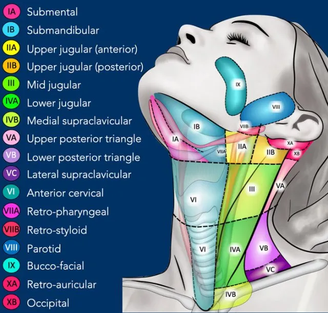
- Neck Triangles: Anterior & Posterior; key subdivisions.
- Lymph Node Levels: I-VII.
- General Classification (📌 CIN):
- Congenital (e.g., thyroglossal, branchial cysts)
- Inflammatory/Infectious (e.g., lymphadenitis)
- Neoplastic (benign/malignant)
- Key History Questions:
- Age, duration, progression (rapid growth?)
- Associated symptoms: Pain, fever, dysphagia, hoarseness, weight loss.
⭐ A persistent, firm, enlarging neck mass in an adult >40 years, especially with smoking/alcohol history, is considered metastatic malignancy until proven otherwise (often SCC from upper aerodigestive tract).
Congenital & Developmental Lumps - Kiddie Knobs
- Thyroglossal Duct Cyst:
- Midline, elevates on tongue protrusion/swallowing.
- Sistrunk procedure for excision.
⭐ Most common congenital neck mass overall.
- Branchial Cleft Cyst:
- Anterior border of SCM.
- Usually smooth, fluctuant; may fistulate.
- 📌 2nd arch most common (Type II).
- Dermoid Cyst:
- Midline (common), can be lateral.
- Doughy consistency, non-tender. Contains adnexal structures.
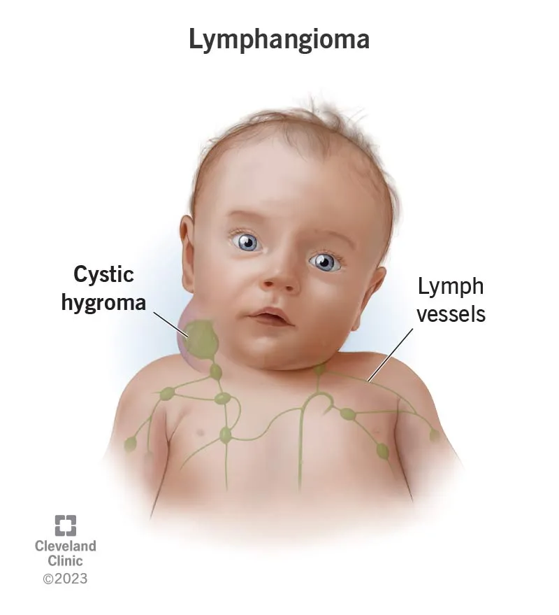
- Cystic Hygroma (Lymphangioma):
- Posterior triangle (classic), can be extensive.
- Soft, compressible, brilliantly transilluminates.
Inflammatory & Infectious Swellings - Fiery Swellings
- Reactive Lymphadenopathy: Most common. Tender, mobile nodes.
- Infectious Lymphadenitis:
- Tuberculous: Scrofuloderma, matted nodes, cold abscess.
⭐ Matted lymph nodes are a classic sign of tuberculosis.
- Bacterial: Staph/Strep. Acute, tender.
- Viral: IMN (EBV), CMV. Posterior triangle common.
- Cat Scratch Disease: Bartonella henselae. Tender nodes post-scratch.
- Tuberculous: Scrofuloderma, matted nodes, cold abscess.
- Deep Neck Space Infections: ⚠️ Airway risk!
- Ludwig's Angina: Submandibular, brawny induration, tongue elevation.
- Parapharyngeal Abscess: Trismus, medial pharyngeal bulge.
Neoplastic Neck Masses - Danger Lumps
- Benign: Lipoma, Fibroma, Neurogenic tumors (Schwannoma, Neurofibroma), Salivary gland pleomorphic adenoma.
- Malignant:
- Metastatic Squamous Cell Carcinoma (SCC): Most common in adults; aerodigestive primary.
- Lymphoma: Hodgkin's & Non-Hodgkin's (B symptoms: fever, night sweats, weight loss).
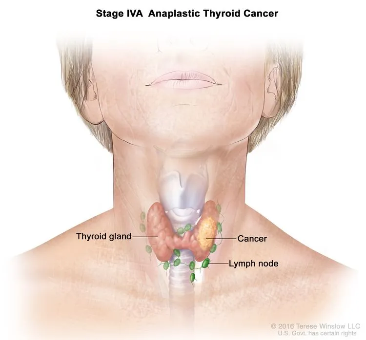
- Thyroid Cancer: Papillary (most common type), follicular, medullary, anaplastic.
- Salivary Gland Malignancies.
- Red Flags ⚠️: Hard, fixed, rapidly growing, age >40 yrs, smoker/drinker history, B symptoms (unexplained fever, night sweats, weight loss), cranial nerve palsies, overlying skin changes (ulceration, discoloration).
⭐ Most common malignant neck mass in adults: Metastatic Squamous Cell Carcinoma, often from an upper aerodigestive tract primary (e.g., oropharynx, larynx).
Diagnostic Pathway - Unraveling Knots
- Clinical Exam: Systematic (Inspect, Palpate: Site, Size, Consistency, Fixity, Nodes).
- Key Investigations:
- USG: Cystic vs. Solid.
- FNAC: Cornerstone for initial tissue diagnosis.
- CT/MRI: Defines extent, relations.
- Biopsy: If FNAC inconclusive.
- Panendoscopy: For suspected occult primary SCC.
⭐ FNAC is the first-line investigation for most neck masses after clinical examination.
High‑Yield Points - ⚡ Biggest Takeaways
- Thyroglossal duct cyst: Most common pediatric midline neck mass; moves with tongue protrusion.
- Branchial cleft cyst: Common pediatric lateral neck mass, anterior to sternocleidomastoid.
- Supraclavicular mass (Virchow's node): Suggests metastatic malignancy from abdomen/thorax.
- Carotid body tumor: Pulsatile lateral neck mass at carotid bifurcation.
- Malignancy suspicion: Firm, fixed, rapidly growing mass in adults.
- Cystic hygroma: Lymphatic malformation, soft, transilluminates, often congenital.
- Tuberculous lymphadenitis: Matted nodes, chronic, may form sinus tract; common in endemic areas like India.
Unlock the full lesson and continue reading
Signup to continue reading this lesson and unlimited access questions, flashcards, AI notes, and more