

Pituitary Gland - Master Controller's Micro-View
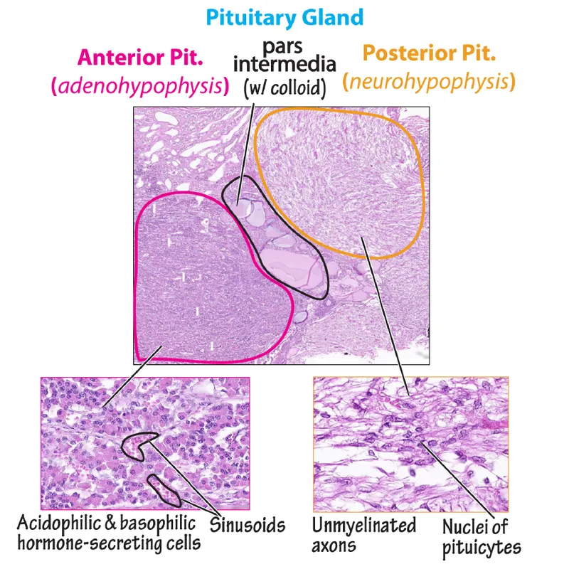
Comprises Adenohypophysis (anterior) & Neurohypophysis (posterior).
- Adenohypophysis (Pars Distalis): Cellular cords & sinusoidal capillaries.
Staining Cells & Hormones Mnemonic Acidophils Somatotrophs (GH), Lactotrophs (PRL) 📌 GPA Basophils Corticotrophs (ACTH), Thyrotrophs (TSH), Gonadotrophs (FSH, LH) 📌 B-FLAT Chromophobes Minimal granules; reserve/degranulated cells. - Pars Intermedia: Basophilic cells (MSH), Rathke's cysts.
- Neurohypophysis (Pars Nervosa):
- Unmyelinated axons from hypothalamus.
- Herring bodies: Axonal dilations with ADH, Oxytocin.
- Pituicytes: Glial support cells.
⭐ Adenohypophysis develops from Rathke's pouch (oral ectoderm); Neurohypophysis from neuroectoderm (diencephalon floor).
Thyroid & Parathyroid - Neck's Hormone Factories
- Thyroid Gland:
- Follicles: Functional units. Lined by simple cuboidal epithelium; lumen contains colloid (thyroglobulin reservoir).
- Parafollicular cells (C cells): Between follicles; secrete calcitonin (regulates $Ca^{2+}$ homeostasis, ↓ serum $Ca^{2+}$).
⭐ Parafollicular cells (C cells) are neural crest derivatives, originating from the ultimobranchial body.
- Parathyroid Glands:
- Chief cells (Principal cells): Predominant; basophilic; secrete Parathyroid Hormone (PTH) (regulates $Ca^{2+}$ homeostasis, ↑ serum $Ca^{2+}$).
- Oxyphil cells: Larger, acidophilic (eosinophilic) cytoplasm; numerous mitochondria; appear around puberty, function uncertain.
 oka
oka
Adrenal Gland - Stress Response Central
- Adrenal Cortex (Zonation & Products):
- Zona Glomerulosa (Outer): Secretes Mineralocorticoids (e.g., Aldosterone $\rightarrow$ Salt balance).
- Zona Fasciculata (Middle, largest): Secretes Glucocorticoids (e.g., Cortisol $\rightarrow$ Sugar metabolism, stress). Cells: Spongiocytes (foamy, lipid-rich).
- Zona Reticularis (Inner): Secretes Androgens (e.g., DHEA $\rightarrow$ Sex characteristics).
- 📌 Mnemonic: GFR (Layers) $\rightarrow$ Salt, Sugar, Sex (Products). "Deeper you go, sweeter it gets."
- Adrenal Medulla (Core):
- Composed of Chromaffin cells (modified postganglionic sympathetic neurons).
- Secretes Catecholamines: Epinephrine (Adrenaline) and Norepinephrine (Noradrenaline) $\rightarrow$ fight-or-flight.

⭐ The Adrenal Medulla is derived from neural crest cells, similar to sympathetic ganglia, and functions as part of the sympathetic nervous system.
Pancreas (Islets) & Pineal - Sugar Boss & Sleep Clock
- Pancreas (Islets of Langerhans): Endocrine cell clusters (1-2% of pancreas mass) regulating glucose.
- Islet Cells & Hormones:
Cell Hormone Location Primary Action Alpha (α) Glucagon Periphery ↑ Blood glucose Beta (β) Insulin Central ↓ Blood glucose Delta (δ) Somatostatin Scattered Inhibits A & B cells PP/Gamma (γ) Pancreatic Polypeptide Periphery Regulates GI/endo - 📌 Insulin INside Beta cells (most numerous).
- Islet Cells & Hormones:
- Pineal Gland: Midline brain structure; melatonin for circadian rhythm.
- Pinealocytes: Chief cells; produce melatonin from serotonin.
- Corpora arenacea (brain sand): Calcified concretions; ↑ with age. No known function.

⭐ Corpora arenacea (brain sand) in the pineal gland are calcified concretions that increase with age and are radiopaque.
High‑Yield Points - ⚡ Biggest Takeaways
- Pituitary: Acidophils (GH, Prolactin), Basophils (FSH, LH, ACTH, TSH); Herring bodies in neurohypophysis.
- Thyroid: Follicles with colloid; Parafollicular C-cells secrete calcitonin.
- Parathyroid: Chief cells (PTH) and larger, eosinophilic Oxyphil cells.
- Adrenal Cortex: Zonation: Glomerulosa (aldosterone), Fasciculata (cortisol), Reticularis (androgens).
- Adrenal Medulla: Chromaffin cells (modified neurons) produce catecholamines.
- Pancreatic Islets: Beta cells (insulin) are central; Alpha cells (glucagon) peripheral.
- Pineal Gland: Pinealocytes (melatonin) and characteristic Corpora arenacea (brain sand).
Unlock the full lesson and continue reading
Signup to continue reading this lesson and unlimited access questions, flashcards, AI notes, and more