

Neurulation - Tube Time Travails
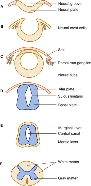
- Notochord induces overlying ectoderm to thicken → neural plate (Day 18).
- Neural plate invaginates centrally → neural groove; lateral edges elevate → neural folds.
- Neural folds fuse dorsally → neural tube (future CNS); fusion starts cervical, extends cranio-caudally.
- Neural crest cells delaminate from lateral edges of neural folds (future PNS, melanocytes).

⭐ Rostral (anterior) neuropore closes by Day 25; failure results in anencephaly. Caudal (posterior) neuropore closes by Day 27-28; failure leads to spina bifida occulta/cystica. Folic acid (Vitamin B9) supplementation pre-conceptionally and in early pregnancy significantly reduces NTD risk.
Brain Vesicles - Brainy Bubbles
- Neural tube's rostral end forms 3 primary vesicles (4th week):
- Prosencephalon (Forebrain)
- Mesencephalon (Midbrain)
- Rhombencephalon (Hindbrain)
- These differentiate into 5 secondary vesicles (5th week):
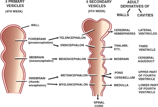
- 📌 Mnemonic (Primary): Prosperous Men Reach. (Prosencephalon, Mesencephalon, Rhombencephalon)
- 📌 Mnemonic (Secondary): Tall Dinos Mess with Metal Myth. (Telencephalon, Diencephalon, Mesencephalon, Metencephalon, Myelencephalon)
⭐ The diencephalon gives rise to the thalamus, hypothalamus, epithalamus, and the retina/optic nerve (via optic vesicles).
Spinal Cord Sprout - Cordial Column
- Origin: Caudal neural tube.
- Neural Tube Wall Layers:
- Ventricular (ependymal): Lines central canal.
- Mantle: Forms grey matter (future horns/columns).
- Alar plates (dorsal): Sensory functions.
- Basal plates (ventral): Motor functions.
- Sulcus limitans: Separates alar/basal.
- Marginal: Becomes white matter.
- Neural Crest Cells → Dorsal Root Ganglia (DRG).
- Relative Ascent: Cord terminates at L1-L2 in adults.
⭐ In adults, the conus medullaris (tapered end of spinal cord) is at L1-L2; lumbar puncture is safely done at L3-L4 or L4-L5.
Neural Crest Cells - Wandering Wonders
- Origin: Neuroectoderm at crest of neural folds; detach during neurulation. Often called the "4th germ layer".
- Migration: Undergo epithelial-mesenchymal transition (EMT) for extensive, guided migration.
- 📌 Key Derivatives:
- PNS: Schwann cells, satellite glial cells, sensory ganglia (DRG), autonomic ganglia, enteric nervous system.
- Craniofacial: Bones, cartilage & CT of face/skull (e.g., Meckel's cartilage), odontoblasts.
- Endocrine/Pigment: Adrenal medulla (chromaffin cells), parafollicular C-cells (thyroid), melanocytes.
- Cardiac: Aorticopulmonary septum, conotruncal & endocardial cushions.
- Meninges: Pia mater & arachnoid mater.
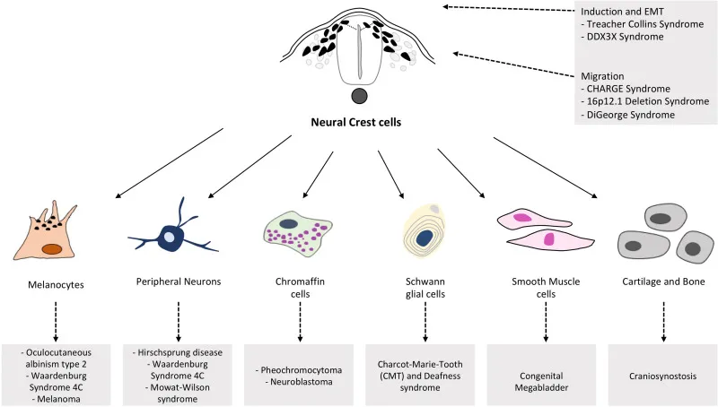
⭐ Key neurocristopathies include Hirschsprung disease (aganglionic megacolon due to failed enteric NCC migration) and DiGeorge syndrome (craniofacial & cardiac defects, often 22q11 deletion).
CNS Malformations - Glitchy Growth
- Neural Tube Defects (NTDs): Failed neural tube closure.
- Anencephaly: Absent forebrain/calvaria.
- Encephalocele: Brain/meninges herniate via skull defect.
- Spina Bifida: Incomplete vertebral arch.
- Occulta: Mild, skin sign (hair tuft).
- Meningocele: Meninges sac.
- Myelomeningocele: Cord + meninges sac; deficits.
- Holoprosencephaly: Failed forebrain cleavage (SHH defects).
- Lissencephaly: "Smooth brain", ↓gyri (migration failure).
- Dandy-Walker: Cystic 4th ventricle, vermis hypoplasia.
- Chiari II: Cerebellar herniation with myelomeningocele.

⭐ Folate (Vit B9) 0.4 mg/day pre-conception & early pregnancy drastically cuts NTD risk.
High‑Yield Points - ⚡ Biggest Takeaways
- Neural tube (CNS) forms from ectodermal neural plate (notochord induction).
- Neural tube defects (anencephaly, spina bifida) from failed closure; folic acid is crucial for prevention.
- Neural crest cells: PNS, melanocytes, adrenal medulla, craniofacial bones.
- Brain vesicles: 3 primary (Pros-, Mes-, Rhombencephalon) expand to 5 secondary.
- Spinal cord: Alar plate (sensory), Basal plate (motor), separated by sulcus limitans.
- Hirschsprung disease: due to failed neural crest cell migration to colon.
Unlock the full lesson and continue reading
Signup to continue reading this lesson and unlimited access questions, flashcards, AI notes, and more