

Somite Saga - Blocks to Body
- Origin: Paired blocks of paraxial mesoderm, appearing craniocaudally from day 20.
- Number: 42-44 pairs form; ~37 pairs persist.
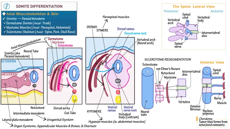
- Differentiation: Each somite differentiates into:
- Sclerotome (ventromedial): Forms axial skeleton (vertebrae, ribs).
⭐ Sclerotomes undergo resegmentation: caudal half of one fuses with cranial half of the next, allowing spinal nerves to pass between vertebrae.
- Dermomyotome (dorsolateral):
- Dermatome: Dermis of the back.
- Myotome: Segmental muscles.
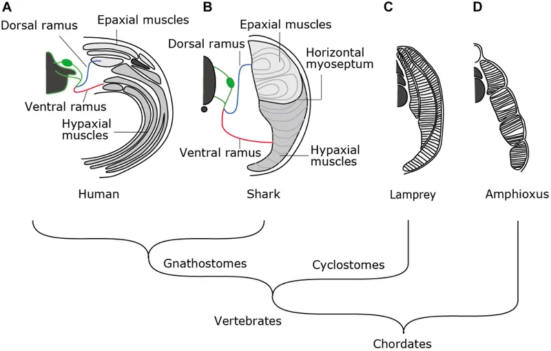
- Epimere (dorsal): Intrinsic back muscles (innervated by dorsal rami).
- Hypomere (ventrolateral): Limb and body wall muscles (innervated by ventral rami).
- Sclerotome (ventromedial): Forms axial skeleton (vertebrae, ribs).
Skeletal Scaffolding - Bones Take Form
- Bone Origins:
- Axial skeleton (vertebrae, ribs): Paraxial mesoderm (sclerotomes).
- Appendicular skeleton (limbs): Lateral plate mesoderm.
- Craniofacial bones: Neural crest cells & paraxial mesoderm.
- Ossification Types:
- Intramembranous: Mesenchyme directly forms bone (e.g., skull flat bones, clavicle).
- Endochondral: Mesenchyme → cartilage model → bone (e.g., long bones, vertebrae, pelvis).
- Primary ossification center (diaphysis).
- Secondary ossification centers (epiphyses) - mostly postnatal.
- 📌 Prenatal secondary centers: Distal femur, proximal tibia (appear before birth).
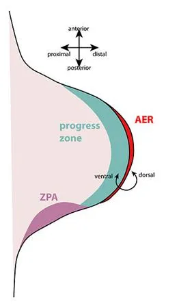
- Limb Development (starts 4th week):
- Apical Ectodermal Ridge (AER): FGFs induce proximo-distal outgrowth.
- Zone of Polarizing Activity (ZPA): Shh signals antero-posterior patterning.
- Dorsal-ventral axis: Wnt7a (dorsal ectoderm), En1 (ventral ectoderm).
- Limb rotation: Upper 90° laterally; lower 90° medially.
- Vertebral Column Formation:
- Sclerotomes resegment: Caudal half of one fuses with cranial half of subjacent sclerotome.
- Intervertebral disc: Notochord → Nucleus pulposus; Sclerotome → Annulus fibrosus.
⭐ Failure of sclerotome resegmentation is a key cause of congenital scoliosis due to vertebral anomalies like hemivertebrae.
Muscle Machine - Making Moves
- Muscles primarily arise from mesoderm.
- Paraxial mesoderm forms somites (42-44 pairs).
- Somites differentiate into:
- Sclerotome (vertebrae, ribs)
- Dermatome (dermis of back)
- Myotome (skeletal muscle)
- Somites differentiate into:
- Myotome divisions:
- Epimere (dorsal): Forms epaxial muscles (e.g., erector spinae). Innervated by dorsal rami of spinal nerves.
- Hypomere (ventrolateral): Forms hypaxial muscles (e.g., limb, abdominal wall muscles). Innervated by ventral rami.
- Myoblasts (from myotomes) fuse, forming multinucleated myotubes → muscle fibers.
- Key myogenic regulatory factor: MyoD.
- Smooth muscle: From splanchnic mesoderm (gut/airways) & somatic mesoderm (blood vessels).
- Cardiac muscle: From splanchnic mesoderm around heart tube.
⭐ Limb musculature develops from the ventrolateral (hypomere) part of somites; cells migrate into the limb bud. These are hypaxial muscles innervated by ventral rami (e.g., brachial plexus, lumbosacral plexus).
Clinical Connections - Development Derails
- Achondroplasia: Most common skeletal dysplasia; impaired endochondral ossification (FGFR3 gene mutation). Results in dwarfism, short limbs, macrocephaly.
- Osteogenesis Imperfecta: Brittle bone disease; defective Type I collagen synthesis (COL1A1/COL1A2 genes). Multiple fractures, blue sclerae, hearing loss.
- Developmental Dysplasia of the Hip (DDH): Abnormal hip joint development; shallow acetabulum, femoral head displacement. Barlow/Ortolani tests.
- Clubfoot (Talipes Equinovarus): Foot twisted out of shape/position. Multifactorial.
- Spina Bifida: Neural tube defect; incomplete vertebral arch closure. Often associated with Chiari II malformation.
⭐ Amniotic Band Syndrome: Can cause constrictions, amputations, or other limb deformities due to entanglement of fetal parts in amniotic bands. Not genetically determined; sporadic occurrence.
High‑Yield Points - ⚡ Biggest Takeaways
- Somites yield sclerotome (vertebrae), myotome (muscle), dermatome (dermis).
- Limb development: AER (FGFs) for proximo-distal axis, ZPA (SHH) for antero-posterior axis.
- HOX genes control segmental patterning of skeleton and limbs.
- Skeletal muscle from paraxial mesoderm; smooth/cardiac from splanchnic mesoderm.
- Bone formation: intramembranous (flat bones) vs. endochondral (long bones).
- Vertebrae from sclerotomes; notochord forms nucleus pulposus.
- Neural crest cells contribute significantly to craniofacial skeleton.
Unlock the full lesson and continue reading
Signup to continue reading this lesson and unlimited access questions, flashcards, AI notes, and more