
Neuroanatomy for Radiologists — Flashcards

The _____ sign is characteristic of superior sagittal sinus thrombosis
_____ view (or occipitofrontal view) is a radiographic view of the skull, where the X-ray plate is angled at 20 to orbitomeatal line.
Ischemic strokes may be visualized on _____ after 6 - 24 hours
Imaging of oligodendroglioma typically reveals a _____ tumor in the white matter, usually involving the frontal lobe
In _____ imaging, CSF is dark/hypointense and white matter is dark, grey matter is white
The _____ sign refers to widely spaced lateral ventricles due to agenesis of the corpus callosum with intervening Probst bundles
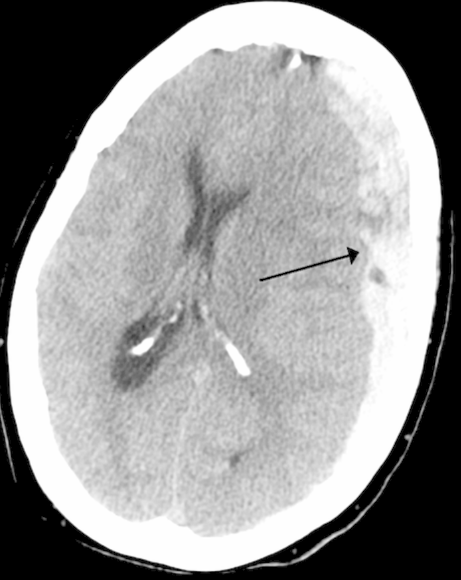
_____ hemorrhage typically occurs in the basal ganglia and internal capsule, but can occur in the cerebral hemispheres, brainstem, and cerebellum
_____ sign on brain CT indicates the presence of tension pneumocephalus.
First Aid Neurology:: Cranial Nerves Pseudotumor Cerebri (aka Idiopathic Intracranial Hypertension) causes a palsy of what oculomotor nerve? _____

CT scan shows a concavo-convex-shaped hematoma is suggestive of a _____ hematoma
Neuroanatomy for Radiologists Flashcards for NEET-PG
Study 10 flashcards on Neuroanatomy for Radiologists for NEET-PG Radiology. These active recall cards cover the key concepts, clinical associations, and high-yield facts from this chapter of Neuroradiology. Each card is designed to test your understanding rather than just recognition, building stronger and more durable memories for exam day.
For personalised spaced repetition scheduling and unlimited flashcards, download the Oncourse app.
Frequently Asked Questions
Are Neuroanatomy for Radiologists flashcards free?
How many flashcards does this chapter have?
How should I use these flashcards for NEET-PG?
Are there more flashcards for Neuroradiology?
Want unlimited flashcards?
Get full access to all flashcards, spaced repetition, and progress tracking.
Start For Free